
Questions
RES001 - Chronic Right Lower Quadrant Mass
Scenario
A 12-year-old boy presents with a 3-month history of low-grade evening fevers, anorexia, significant weight loss (4 kg), and intermittent postprandial cramping right lower quadrant pain. Physical examination reveals pallor, mild abdominal distension, and a firm, non-tender, ill-defined fullness palpated in the right iliac fossa. Contrast-enhanced computed tomography (CECT) of the abdomen and ultrasonography are performed to evaluate for suspected ileocecal pathology.
Questions
- Enumerate five classical contrast-enhanced CT (CECT) or ultrasonographic abdominal imaging features characteristic of ileocecal tuberculosis.
- What specific lymph node morphology on contrast-enhanced CT reliably differentiates this condition from intestinal lymphoma?
- Contrast three structural characteristics of the bowel strictures observed in this disease entity with those seen in Crohn's disease.
- Specify the first-line antitubercular therapy (ATT) regimen, including weight-based daily drug doses and total treatment duration, as per the National Tuberculosis Elimination Program (NTEP) guidelines.
Answer
- Classical CECT / USG Findings:
- Concentric, circumferential, symmetric mural thickening involving the terminal ileum, ileocecal valve, and medial cecal wall.
- Contracted, shrunken, and pulled-up cecum situated abnormally high in the lumbar or subhepatic region, with loss of the normal acute ileocecal angle (obtuse, flattened angle).
- Multiple, short-segment (<3 cm), smooth strictures with symmetrical upstream luminal dilatation.
- Mesenteric and retroperitoneal lymphadenopathy characterized by central non-enhancing hypodensity (caseation necrosis) and peripheral rim enhancement.
- High-attenuation or loculated ascites with fine internal fibrinous septations, peritoneal thickening, and engorgement of adjacent mesenteric vessels ("comb sign").
- Differentiating Lymph Node Morphology:
- Tuberculous lymph nodes exhibit central caseous necrosis (hypoattenuation <20 Hounsfield Units) with peripheral rim enhancement.
- Lymphomatous nodes demonstrate homogeneous enhancement without central necrosis (prior to chemotherapy) and typically form large, confluent, lobulated retroperitoneal nodal masses that encase vessels without luminal compression ("sandwich sign").
- Stricture Characteristics: Intestinal Tuberculosis vs. Crohn's Disease:
- Length: Tuberculous strictures are typically short-segment (<3 cm), whereas Crohn's strictures are longer (>5–10 cm).
- Distribution: Tuberculous strictures are commonly multiple and concentric; Crohn's disease demonstrates asymmetric, transmural, eccentric involvement with skipped segments.
- Associated Signs: Tuberculosis lacks extensive fibrofatty mesenteric proliferation; Crohn's disease prominently exhibits mesenteric fat wrapping ("creeping fat") and deep transmural fissuring ulcers with fistulae.
- NTEP Pediatric Treatment Protocol:
- Intensive Phase (2 months - HRZE daily):
- Isoniazid (H): $10\text{ mg/kg/day}$ (range: 7–15 mg/kg/day, maximum 300 mg/day)
- Rifampicin (R): $15\text{ mg/kg/day}$ (range: 10–20 mg/kg/day, maximum 600 mg/day)
- Pyrazinamide (Z): $35\text{ mg/kg/day}$ (range: 30–40 mg/kg/day, maximum 2000 mg/day)
- Ethambutol (E): $20\text{ mg/kg/day}$ (range: 15–25 mg/kg/day, maximum 1200 mg/day)
- Continuation Phase (4 months - HRE daily):
- Isoniazid (H), Rifampicin (R), and Ethambutol (E) at the same weight-band daily doses.
- Total Duration: 6 months (may be extended to 9–12 months only in cases of slow clinical resolution or extensive multi-organ involvement).
- Intensive Phase (2 months - HRZE daily):
RES002 - Adolescent Respiratory Distress Blood Gas
Scenario
A 14-year-old girl with severe kyphoscoliosis presents to the emergency pediatric intensive care unit with acute-on-chronic respiratory distress precipitated by an intercurrent viral infection. She is receiving supplemental oxygen via a high-flow Venturi mask delivering a fraction of inspired oxygen ($\text{FiO}_2$) of $0.60$ at sea level ($\text{P}_{\text{atm}} = 760\text{ mmHg}$, water vapor pressure $\text{P}_{\text{H}_2\text{O}} = 47\text{ mmHg}$).
An arterial blood gas (ABG) sample drawn on $\text{FiO}_2\ 0.60$ reveals:
- $\text{pH}: 7.34$
- $\text{PaCO}_2: 60\text{ mmHg}$
- $\text{PaO}_2: 48\text{ mmHg}$
- $\text{HCO}_3^-: 32\text{ mEq/L}$
- $\text{SaO}_2: 82\%$
Questions
- Provide a step-by-step diagnostic interpretation of this arterial blood gas (primary acid-base status, degree of compensation, and oxygenation state).
- Calculate the alveolar-arterial oxygen gradient ($\text{A-a DO}_2$) using the standard Alveolar Gas Equation (assume Respiratory Quotient $\text{R} = 0.8$). Show all mathematical derivation steps.
- State the expected normal reference range for the $\text{A-a DO}_2$ on room air in a healthy child, and explain the pathophysiological significance of this patient's calculated gradient.
- Outline two critical non-invasive or invasive positive-pressure ventilatory strategies indicated to treat this patient's dual gas-exchange derangements.
Answer
- Systematic Blood Gas Interpretation:
- Acid-Base Balance: $\text{pH}\ 7.34$ reflects mild uncorrected acidemia ($\text{pH} < 7.35$).
- Primary Disturbance: $\text{PaCO}_2\ 60\text{ mmHg}$ (elevated $>45\text{ mmHg}$) indicates primary respiratory acidosis.
- Compensatory Response: $\text{HCO}_3^-\ 32\text{ mEq/L}$ (elevated $>26\text{ mEq/L}$). For chronic respiratory acidosis, expected $\Delta\text{HCO}_3^- = 0.35\text{ to }0.40 \times \Delta\text{PaCO}_2$. Here, $\Delta\text{PaCO}_2 = 60 - 40 = 20\text{ mmHg}$. Expected $\text{HCO}_3^- = 24 + (20 \times 0.35\text{–}0.40) = 31\text{–}32\text{ mEq/L}$. The measured bicarbonate ($32\text{ mEq/L}$) matches this expected response, confirming a partially compensated chronic respiratory acidosis (or chronic hypercapnia with acute worsening).
- Oxygenation: $\text{PaO}_2\ 48\text{ mmHg}$ on an $\text{FiO}_2$ of $0.60$ indicates severe hypoxemic respiratory failure ($\text{P/F ratio} = 48 / 0.60 = 80$).
- Alveolar-Arterial Oxygen Difference ($\text{A-a DO}_2$) Calculation: $$\begin{aligned} \text{PAO}_2 &= \left[(\text{P}_{\text{atm}} - \text{P}_{\text{H}_2\text{O}}) \times \text{FiO}_2\right] - \frac{\text{PaCO}_2}{\text{R}} \\ &= \left[(760 - 47) \times 0.60\right] - \frac{60}{0.8} \\ &= (713 \times 0.60) - 75 \\ &= 427.8 - 75 = \mathbf{352.8\text{ mmHg}} \\ \text{A-a DO}_2 &= \text{PAO}_2 - \text{PaO}_2 \\ &= 352.8 - 48 \\ &= \mathbf{304.8\text{ mmHg}} \quad (\text{if using } \text{R}=1.0\text{: } 427.8 - 60 - 48 = \mathbf{319.8\text{ mmHg}}) \end{aligned} $$
- Normal Range and Pathophysiological Significance:
- Normal Range: On room air ($\text{FiO}_2\ 0.21$), normal $\text{A-a DO}_2$ is $<10\text{–}15\text{ mmHg}$ (or estimated as $\frac{\text{Age in years}}{4} + 4$). On $\text{FiO}_2\ 0.60$, normal $\text{A-a DO}_2$ is generally $<100\text{ mmHg}$.
- Significance: A markedly widened gradient ($>300\text{ mmHg}$) proves that hypoxemia is not solely due to extrapulmonary alveolar hypoventilation (from chest wall deformity). It confirms significant intrinsic intrapulmonary pathology causing ventilation-perfusion ($\text{V/Q}$) mismatch, atelectasis, right-to-left intrapulmonary shunt, or alveolar consolidation.
- Ventilatory Management Priorities:
- Augment Alveolar Ventilation (Address Hypercapnia): Initiate Non-Invasive Positive Pressure Ventilation (BiPAP) or invasive mechanical ventilation with adequate driving pressure / pressure support ($\Delta P = \text{IPAP} - \text{EPAP}$) and tidal volumes ($6\text{–}8\text{ mL/kg}$) to increase minute ventilation and clear accumulated $\text{CO}_2$.
- Recruit Atelectatic Lung & Optimize Oxygenation: Apply positive end-expiratory pressure (PEEP / EPAP $6\text{–}10\text{ cmH}_2\text{O}$) to recruit collapsed dependent alveoli, improve functional residual capacity (FRC), minimize intrapulmonary shunting, and allow weaning of toxic $\text{FiO}_2$.
RES003 - Recurrent Aspiration and Regurgitation Evaluation
Scenario
An 18-month-old girl is referred to pediatric pulmonology due to three episodes of right middle and lower lobe pneumonia over the past 8 months, requiring repeated hospitalizations. Her mother reports that since 6 months of age, she has had persistent effortless regurgitation of undigested, non-bilious, non-acidic food ingested hours earlier, especially when placed supine at night. She has poor weight gain (weight-for-age $<3\text{rd}$ percentile). A timed barium swallow examination is obtained.
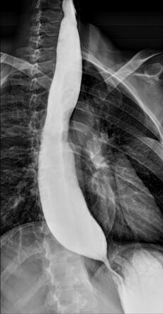 (
Image Source
)
Questions
- Identify the radiological investigation performed and describe the two classical diagnostic imaging findings seen at the gastroesophageal junction and esophageal body.
- Name the gold standard diagnostic investigation for confirming this condition and state the pathognomonic diagnostic manometric criteria.
- State the underlying neuro-histopathological defect responsible for this disorder.
- Detail the definitive medical and surgical therapeutic modalities available for pediatric management.
Answer
Radiological Investigation & Classical Findings:
- Investigation: Barium Swallow / Barium Esophagogram.
- Key Findings:
- Distal Esophagus: Smooth, symmetric, conical/beak-like tapering at the gastroesophageal junction ("bird's-beak" or "rat-tail" appearance).
- Esophageal Body: Marked dilatation of the thoracic esophagus ("megaesophagus") with absent primary peristalsis, retained food/fluid debris, and delayed passage of barium contrast into the gastric fundus.
Gold Standard Investigation & Manometric Criteria:
- Investigation: High-Resolution Esophageal Manometry (HRM).
- Diagnostic Criteria (Chicago Classification):
- Elevated Integrated Relaxation Pressure (IRP; $>15\text{ mmHg}$), indicating incomplete or absent relaxation of the lower esophageal sphincter (LES) during swallowing.
- Aperistalsis in the esophageal tubular body (absence of normal coordinated peristaltic contractions; classified into Type I classic aperistalsis, Type II with panesophageal pressurization, or Type III spastic achalasia).
Neuro-Histopathological Defect:
- Progressive autoimmune or degenerative inflammatory loss and degeneration of inhibitory ganglion cells (nitrinergic and VIPergic neurons) in the myenteric (Auerbach's) plexus of the distal esophagus and LES.
- Unopposed excitatory cholinergic innervation results in impaired nitric oxide/VIP release, leading to sustained tonic contraction/incomplete relaxation of the LES and aperistalsis.
Definitive Therapeutic Modalities:
- Definitive Surgical Intervention:
- Laparoscopic Heller Cardiomyotomy: Longitudinal myotomy dividing the circular muscle layers of the distal esophagus (extending $5\text{–}6\text{ cm}$ above the gastroesophageal junction) and proximal stomach ($2\text{ cm}$ below).
- Antireflux Procedure: Routine partial fundoplication (anterior Dor or posterior Toupet partial wrap) is performed concurrently to prevent debilitating postoperative gastroesophageal reflux.
- Endoscopic Interventions:
- Per-Oral Endoscopic Myotomy (POEM): Minimally invasive submucosal tunnel myotomy of the inner circular muscle layer; highly effective alternative in older children and adolescents.
- Pneumatic Balloon Dilatation (PD): Graded balloon dilation ($30\text{ mm}$, $35\text{ mm}$) to forcibly tear LES circular fibers; carries higher risk of perforation ($1\text{–}3\%$) and recurrent symptoms in young children.
- Medical / Temporizing Options:
- Smooth muscle relaxants: Calcium channel blockers (oral Nifedipine $0.2\text{–}0.5\text{ mg/kg}$ 30 min before meals) or sublingual nitrates.
- Endoscopic Botulinum toxin (Botox) injection into the LES (short-term temporizing measure reserved for high surgical risk or before definitive intervention).
- Definitive Surgical Intervention:
RES004 - Persistent Nasal Discharge And Headache
Scenario
An 8-year-old boy weighing 24 kg is brought to the pediatric outpatient clinic with a 10-day history of continuous nasal congestion and thick rhinorrhea, accompanied by a persistent daytime cough. Over the preceding 3 days, he developed worsening bilateral facial aching, frontal headache, and intermittent low-grade fever (38.1°C / 100.6°F) that did not respond significantly to oral paracetamol. His parents note that his symptoms initially seemed like a standard cold but progressively worsened rather than resolving.
On examination, his temperature is 38.0°C, pulse rate 92/min, respiratory rate 20/min, and blood pressure 104/66 mmHg. Anterior rhinoscopy demonstrates hyperemic, edematous nasal mucosa with thick, purulent discharge visible along the middle meatus bilaterally. There is prominent tenderness upon digital percussion over both maxillary prominences. Visual acuity is normal, ocular movements are full and painless, and no periorbital swelling or meningeal signs are present.
Questions
- Enumerate the three distinct clinical presentation patterns recognized by pediatric guidelines (e.g., American Academy of Pediatrics) for establishing a clinical diagnosis of acute bacterial rhinosinusitis without imaging.
- Identify the three most common bacterial etiologies implicated in community-acquired acute bacterial rhinosinusitis in children.
- What is the recommended role of routine plain sinus radiography or computed tomography (CT) in uncomplicated acute bacterial rhinosinusitis? State one clear clinical indication for urgent contrast-enhanced CT of the orbits and paranasal sinuses.
- Calculate the recommended first-line oral antimicrobial regimen and exact daily dose (mg/day and dosing interval) for this 24 kg child in a geographic area with low rates of penicillin-resistant pathogens.
- If the child has persistent fever and worsening facial pain after 72 hours of compliant first-line therapy, what is the next step in antimicrobial management? Provide the drug formulation and weight-based dosage.
- List four "red flag" clinical findings that indicate immediate evaluation for suppurative orbital or intracranial complications.
Answer
AAP Clinical Presentation Patterns for Acute Bacterial Rhinosinusitis:
- Persistent illness: Nasal discharge (of any consistency: serous, mucoid, or purulent) or daytime cough (or both) lasting for $\ge 10$ days without any clinical improvement.
- Worsening course ("double sickening"): Worsening or new onset of nasal discharge, daytime cough, or new fever following a period of initial clinical improvement from a typical viral upper respiratory infection (typically around day 6 to 7).
- Severe onset: Concurrent high fever ($\ge 39^\circ\text{C}$ or $102.2^\circ\text{F}$) accompanied by purulent nasal discharge for at least 3 consecutive days at the onset of illness.
Causative Bacterial Pathogens:
- Streptococcus pneumoniae (30% to 40%)
- Non-typeable Haemophilus influenzae (30% to 40%)
- Moraxella catarrhalis (10% to 15%)
Role of Diagnostic Imaging & Urgent Indication:
- Plain radiography: Not recommended; poor sensitivity and specificity, inability to differentiate mucosal thickening or air-fluid levels of viral viral upper respiratory infections from bacterial disease.
- Computed tomography (CT): Not recommended for routine, uncomplicated acute bacterial rhinosinusitis due to high cost and unnecessary ionizing radiation.
- Indication for urgent contrast-enhanced CT: Clinical suspicion of orbital or intracranial extension (e.g., proptosis, ophthalmoplegia, visual impairment, severe periorbital edema, focal neurological signs, meningismus, or severe refractory headache).
First-Line Antimicrobial Dosing:
- Agent: Oral Amoxicillin (standard dose) or Amoxicillin-Clavulanate.
- Calculation: $$\begin{aligned} \text{Daily Dose} &= 45\text{ mg/kg/day} \times 24\text{ kg} = 1080\text{ mg/day} \\ \text{Divided Regimen} &= 500\text{ mg PO twice daily (every 12 hours) for 10 days} \end{aligned} $$ (Note: If high-dose amoxicillin is selected for endemic resistance: $90\text{ mg/kg/day} = 2160\text{ mg/day}$, administered as $1000\text{ mg}$ PO twice daily).
Second-Line Antimicrobial Management (Treatment Failure at 48–72 Hours):
- Agent: High-dose Amoxicillin-Clavulanate ($14:1$ formulation to minimize clavulanate-induced diarrhea).
- Dose: $90\text{ mg/kg/day}$ of the amoxicillin component with $6.4\text{ mg/kg/day}$ of clavulanate orally divided every 12 hours.
- Calculation for 24 kg child: $$\begin{aligned} \text{Amoxicillin Component} &= 90\text{ mg/kg/day} \times 24\text{ kg} = 2160\text{ mg/day} \\ \text{Administered Dose} &= 1000\text{ mg PO twice daily (every 12 hours) for 10 days} \end{aligned} $$ (Alternative for patients unable to tolerate oral medications: Ceftriaxone $50\text{ mg/kg/day}$ IV/IM once daily for 1 to 3 days, followed by oral step-down once improved).
Red Flag Clinical Signs for Complications:
- Proptosis or exophthalmos
- Ophthalmoplegia (restricted or painful extraocular muscle movement)
- Decreased visual acuity, diplopia, or loss of color vision
- Marked periorbital edema, erythema, or chemosis extending beyond the orbital rim
- Altered mental status, severe photophobia, or meningismus
- Fluctuant forehead soft tissue swelling (Pott's puffy tumor due to subperiosteal abscess of the frontal bone)
More Details
Clinical Pearl: Differentiating Viral URI from Bacterial Rhinosinusitis
An uncomplicated viral rhinosinusitis typically peaks in symptom severity between days 3 and 5 and resolves or substantially improves within 7 to 10 days. The diagnosis of acute bacterial rhinosinusitis (ABRS) is strictly clinical; routine sinus imaging (plain X-rays or CT) fails to discriminate between viral mucosal edema and bacterial pus.
flowchart TD
    A["Suspected Acute Rhinosinusitis Presentation"] --> B{"Clinical Presentation Pattern"}
    B -->|"Persistent: Nasal discharge / daytime cough ≥10 days without clinical improvement"| C["Acute Bacterial Rhinosinusitis (ABRS)"]
    B -->|"Double Sickening: Initial URI improvement, then abrupt worsening fever/discharge at day 6–7"| C
    B -->|"Severe: High fever (≥39°C) + purulent nasal discharge for ≥3 consecutive days"| C
    B -->|"Mild to moderate symptoms improving steadily within 7–10 days"| D["Viral Upper Respiratory Tract Infection (URI)"]
Chandler Classification of Orbital Complications of Sinusitis
- Group I: Preseptal cellulitis (edema anterior to the orbital septum, normal ocular motility and acuity)
- Group II: Orbital cellulitis (diffuse post-septal infiltration, chemosis, proptosis, no discrete collection)
- Group III: Subperiosteal abscess (collection between periorbita and bony orbital wall, displacing the globe)
- Group IV: Orbital abscess (discrete purulent collection within the intraconal or extraconal fat, severe ophthalmoplegia and vision loss risk)
- Group V: Cavernous sinus thrombosis (bilateral orbital symptoms, cranial neuropathies III, IV, V1, V2, VI, rapid progression, high mortality)
RES005 - Infant Wheezing And Respiratory Distress
Scenario
A 7-month-old male infant is brought to the pediatric emergency department during winter with a 3-day history of clear rhinorrhea, low-grade fever, and progressive difficulty in breathing. On examination, he is alert but irritable, with tachypnea and feeding refusal.
Vital signs:
- Heart rate: 138/min
- Respiratory rate: 70/min
- Temperature: 37.6°C
- Oxygen saturation ($SpO_2$): 88% on ambient air
- Capillary refill time: <2 seconds
Respiratory examination reveals nasal flaring, moderate intercostal and subcostal recessions, an increased anteroposterior diameter of the thorax, and liver dullness pushed down to 2 cm below the right costal margin. Auscultation demonstrates prolonged expiration, bilateral widespread fine inspiratory crackles (crepitations), and high-pitched polyphonic expiratory wheezing throughout both lung fields.
Questions
- What is the most likely clinical diagnosis?
- Name the primary viral pathogen responsible for >50–70% of these cases, and list three other recognized viral causative agents.
- List four high-risk host factors that predispose an infant with this condition to severe disease, respiratory failure, or apnea.
- Outline the primary evidence-based supportive management in the acute emergency setting, and list three pharmacological interventions routinely not recommended by international consensus guidelines (AAP / NICE).
Answer
Most Likely Clinical Diagnosis:
- Acute Bronchiolitis (first episode of acute wheezing preceded by viral upper respiratory prodrome in an infant <12 months of age).
Etiological Pathogens:
- Primary pathogen (>50–70%): Respiratory Syncytial Virus (RSV; Orthopneumovirus, Subgroups A and B).
- Other causative agents (any three):
- Human Rhinovirus (HRV)
- Human Metapneumovirus (hMPV)
- Parainfluenza virus (predominantly type 3)
- Adenovirus
- Human Bocavirus
- Influenza virus (A and B)
- Seasonal Coronaviruses
High-Risk Host Factors for Severe Disease / Apnea:
- Prematurity (gestational age <37 weeks, especially <32 weeks).
- Young chronological age at presentation (<6 to 12 weeks of life).
- Hemodynamically significant congenital heart disease (e.g., cyanotic defects, left-to-right shunts with pulmonary hypertension).
- Chronic lung disease of prematurity (bronchopulmonary dysplasia).
- Congenital or acquired immunodeficiency syndromes.
- Underlying neuromuscular disorders or anatomical airway abnormalities.
Management & Non-Recommended Therapies:
- Indicated Evidence-Based Supportive Care:
- Supplemental Oxygen: Humidified oxygen via low-flow nasal cannula or heated humidified high-flow nasal cannula (HFNC) titrated to maintain $SpO_2 \ge 90\text{--}92\%$.
- Respiratory Support: Trial of HFNC (1–2 L/kg/min) or CPAP if severe work of breathing or impending respiratory failure occurs.
- Fluid & Hydration Maintenance: Nasogastric/orogastric enteral feeding (fractionated small feeds) if safe; switch to intravenous isotonic fluids ($0.9\%\text{ NaCl in }5\%\text{ Dextrose}$) at maintenance rates if respiratory rate >60–70 breaths/min or aspiration risk is high.
- Airway Clearance: Gentle superficial upper airway/nasal suctioning before feeds and sleep.
- Routinely NOT Recommended (AAP / NICE Guidelines):
- Systemic or inhaled corticosteroids (e.g., oral prednisolone, IV hydrocortisone, inhaled budesonide).
- Routine antibacterial agents (unless concurrent secondary bacterial infection like acute otitis media or severe sepsis is documented).
- Routine inhaled bronchodilators (e.g., salbutamol, ipratropium).
- Routine nebulized hypertonic saline in the acute emergency department setting.
- Routine chest physiotherapy.
- Indicated Evidence-Based Supportive Care:
More Details
- Necrosis and sloughing of respiratory epithelial cells into the lumen.
- Peribronchiolar lymphocytic and mononuclear cellular infiltration with subepithelial edema.
- Hypersecretion of mucus forming plugs with fibrin and cellular debris.
Because airway resistance is inversely proportional to the fourth power of the radius ($R \propto 1/r^4$ according to Poiseuille's law), even minor luminal narrowing causes severe, disproportionate airway obstruction, dynamic hyperinflation (ball-valve effect), and patchy atelectasis resulting in ventilation-perfusion ($\dot{V}/\dot{Q}$) mismatch and hypoxemia. Bronchospasm plays a negligible role in infants <12 months, which explains why $\beta_2$-agonists are clinically ineffective.
RES006 - Sickle Cell Thoracic Distress
Scenario
A 12-year-old boy with homozygous sickle cell disease (HbSS), weighing 32 kg, presents to the pediatric emergency department during winter with a 6-hour history of severe right-sided chest pain, bilateral tibial bone pain, and high-grade fever. On examination, he is anxious, pale, and tachypneic. Vital signs: temperature 39.4°C, heart rate 138 beats/min, respiratory rate 46 breaths/min, blood pressure 104/66 mmHg, and $\text{SpO}_2\text{ 84\%}$ on room air. Chest examination reveals intercostal retractions, dullness to percussion over the right lower hemithorax, and coarse crepitations with bronchial breath sounds over the right infrascapular region.
Questions
- State the most likely diagnosis and its definitive diagnostic criteria.
- Outline four major underlying pathophysiological mechanisms contributing to this pulmonary event.
- Formulate the criteria for simple blood transfusion versus emergent automated red blood cell exchange transfusion (erythrocytapheresis) in this setting.
- Detail the acute management plan, specifying weight-based analgesia, empirical antimicrobial therapy, and preventive respiratory supportive care.
Answer
- Diagnosis & Diagnostic Criteria:
- Diagnosis: Acute Chest Syndrome (ACS) complicating Sickle Cell Disease.
- Criteria: A new pulmonary segmental or lobar alveolar infiltrate on chest radiography involving at least one complete lung segment (typically lower lobes), accompanied by at least one of the following:
- Temperature $\ge 38.5^\circ\text{C}$
- Tachypnea, intercostal retractions, or chest pain
- Hypoxemia ($\gt 3\%$ absolute decrease from baseline $\text{SpO}_2$ or room air $\text{SpO}_2 \lt 90\%$)
- Auscultatory findings (crackles, wheezing, or bronchial breathing)
- Pathophysiological Mechanisms:
- Pulmonary microvascular vaso-occlusion: Entrapment and adhesion of sickled erythrocytes to vascular endothelium leading to regional ischemia and infarction.
- Infection: Atypical organisms (Mycoplasma pneumoniae, Chlamydophila pneumoniae) and encapsulated bacteria (Streptococcus pneumoniae) trigger local inflammation, hypoxemia, and secondary sickling.
- Fat and bone marrow embolism: Necrosis of hematopoietic bone marrow during painful vaso-occlusive crisis releases marrow fat globules into the venous circulation, lodging in pulmonary microvasculature.
- Hypoventilation and atelectasis: Severe thoracic or abdominal wall bone pain leads to splinting, shallow breathing, regional atelectasis, and worsened ventilation-perfusion mismatch.
- Transfusion Indications:
- Simple Blood Transfusion (Packed Red Blood Cells, 10 mL/kg):
- Indicated in mild-to-moderate ACS with an acute drop in hemoglobin $\gt 1\text{--}2\text{ g/dL}$ below baseline, provided the post-transfusion hemoglobin does not exceed $10\text{--}11\text{ g/dL}$ (to prevent hyperviscosity crisis).
- Emergent Exchange Transfusion (Erythrocytapheresis):
- Indicated in rapidly progressive or severe ACS: $\text{PaO}_2 \lt 60\text{ mmHg}$, $\text{SpO}_2 \lt 90\%$ despite high-flow supplemental oxygen, multi-lobar pulmonary involvement, hemodynamic instability, or deteriorating neurological status.
- Target: Rapid reduction of $\text{HbS} \lt 30\%$ while maintaining total hemoglobin between $10\text{ and }11\text{ g/dL}$.
- Simple Blood Transfusion (Packed Red Blood Cells, 10 mL/kg):
- Acute Management & Dosing:
- Oxygenation & Supportive Care: Maintain $\text{SpO}_2 \ge 95\%$ using humidified high-flow nasal cannula or non-invasive positive pressure ventilation (NIV/CPAP); judicious IV hydration at maintenance rate ($100\%$ maintenance; avoid overhydration to prevent pulmonary edema).
- Analgesia:
- IV Morphine: $0.1\text{ mg/kg/dose}$ IV every 3–4 hours PRN (or patient-controlled analgesia; $32\text{ kg} \times 0.1\text{ mg/kg} = \mathbf{3.2\text{ mg/dose}}$), titrated carefully with respiratory monitoring.
- Empirical Antimicrobial Therapy:
- IV Ceftriaxone: $75\text{--}100\text{ mg/kg/day}$ IV once daily or divided q12h (max $4\text{ g/day}$; for $32\text{ kg} = \mathbf{2.4\text{ to }3.2\text{ g/day}}$).
- PLUS Oral/IV Azithromycin: $10\text{ mg/kg/day}$ once daily (max $500\text{ mg/day}$; for $32\text{ kg} = \mathbf{320\text{ mg/day}}$) to cover atypical pathogens.
- Respiratory Support / Prevention:
- Incentive spirometry: 10 breaths every 2 hours while awake (proven to prevent ACS progression during vaso-occlusive bone crisis).
RES007 - Acute Febrile Upper Airway Obstruction
Scenario
A 4-year-old unimmunized girl weighing 16 kg presents to the pediatric emergency resuscitation bay with a 10-hour history of high-grade fever, sore throat, and rapidly progressive breathing difficulty. The child appears toxic, pale, and markedly anxious. She is sitting upright, leaning forward with her neck extended, mouth open, and chin thrust forward, with noticeable drooling of secretions. Auscultation reveals a soft, low-pitched inspiratory stridor with prominent sternal and intercostal retractions. A lateral soft-tissue radiograph of the neck is performed under physician supervision.
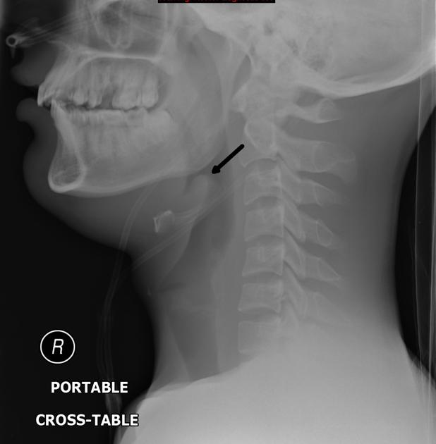 (
Image Source
)
Questions
- Identify the clinical posture demonstrated by the child and the classic radiological sign visualized on the lateral neck radiograph.
- What is the most likely clinical diagnosis, and what is the definitive diagnostic method including critical procedural precautions?
- Enumerate three microbiological pathogens most frequently implicated in this condition.
- Detail the immediate emergency airway intervention and empirical weight-based parenteral antimicrobial therapy.
Answer
- Clinical Posture & Radiological Sign:
- Clinical Posture: "Tripod position" (or "sniffing dog position") — upright posture, trunk leaning forward, resting on hands with neck hyperextended, chin projected outward, and mouth open to maximize airway caliber.
- Radiological Sign: "Thumbprint sign" (or "Thumb sign") — rounded, swollen, thickened epiglottis mimicking a thumb protruding into the hypopharynx with obliteration of the vallecular air space.
- Diagnosis & Definitive Diagnostic Precautions:
- Diagnosis: Acute Epiglottitis (Supraglottitis).
- Definitive Diagnostic Method: Direct visualization of an edematous, cherry-red, swollen epiglottis and arytenoepiglottic folds via direct laryngoscopy or fiberoptic nasopharyngolaryngoscopy.
- Procedural Precautions:
- Must ONLY be performed in a fully equipped operating room or intensive care unit.
- Must have personnel skilled in pediatric advanced airway management present (pediatric anesthesiologist and ENT surgeon).
- Emergency surgical airway equipment (rigid bronchoscope, cricothyroidotomy, tracheostomy tray) immediately accessible and open.
- Avoid invasive oral examination with a tongue depressor or pharyngeal swabs in the emergency department, as it can precipitate fatal acute laryngospasm.
- Causative Microorganisms:
- Haemophilus influenzae type b (Hib) (predominant in unimmunized/incompletely immunized individuals).
- Streptococcus pyogenes (Group A $\beta$-hemolytic Streptococcus).
- Streptococcus pneumoniae.
- Staphylococcus aureus (including Methicillin-resistant S. aureus [MRSA]).
- Emergency Airway Intervention & Antimicrobial Therapy:
- Airway Intervention:
- Minimize agitation; keep the child comfortably in the parent’s lap.
- Transfer immediately to the operating room for smooth inhalational induction (e.g., sevoflurane in $100\%\ \text{O}_2$) maintaining spontaneous respiration, followed by endotracheal intubation using an uncuffed or cuffed tube $0.5\text{--}1.0\text{ mm}$ smaller than the age-predicted size.
- Antimicrobial Regimen:
- IV Ceftriaxone: $100\text{ mg/kg/day}$ IV once daily or divided q12h (for $16\text{ kg} = \mathbf{1600\text{ mg/day}}$ or $800\text{ mg}$ IV q12h; max $4\text{ g/day}$).
- PLUS IV Vancomycin (if MRSA or resistant Streptococcus suspected): $40\text{--}60\text{ mg/kg/day}$ divided q6h (for $16\text{ kg} = 160\text{--}240\text{ mg}$ IV every 6 hours).
- Continue IV therapy for 7–10 days, transitioning to oral therapy once extubated and clinically afebrile for 48–72 hours.
- Airway Intervention:
RES008 - Pediatric Cough And Tuberculosis Contact
Scenario
An 8-month-old male infant weighing 8.0 kg is brought by his parents to the pediatric outpatient department for evaluation. The family recently discovered that their resident domestic caregiver was diagnosed with microbiologically confirmed, sputum smear-positive (3+) pulmonary tuberculosis 1 week ago. The infant has had a mild dry cough and serous rhinorrhea for 2 days without fever. The infant is alert, feeding well, and has no respiratory distress. Vital signs: heart rate 124 beats/min, respiratory rate 38 breaths/min, and $\text{SpO}_2\text{ 98\%}$ on room air.
Questions
- Enumerate the age-specific respiratory rate cut-offs defining tachypnea under the WHO/IMNCI Integrated ARI guidelines, and list four cardinal clinical signs that classify a child under "Severe Pneumonia or Very Severe Disease".
- Outline the systematic diagnostic algorithm required to exclude active tuberculosis in this 8-month-old exposed infant before starting preventive therapy.
- Calculate the exact daily dose and total duration of Isoniazid preventive therapy (IPT) for this infant according to the National Tuberculosis Elimination Program (NTEP) / WHO guidelines.
- State the indication, clinical rationale, and exact dosing of Pyridoxine (Vitamin B6) co-administration during this child's chemoprophylaxis.
Answer
- IMNCI ARI Tachypnea Cut-offs & Severe Pneumonia Signs:
- Age-specific tachypnea thresholds:
- Age $\lt 2\text{ months}$: $\ge 60\text{ breaths/min}$
- Age $2\text{ to }11\text{ months}$: $\ge 50\text{ breaths/min}$
- Age $12\text{ to }59\text{ months}$: $\ge 40\text{ breaths/min}$
- Signs of Severe Pneumonia / Very Severe Disease (any one of the following):
- Lower chest indrawing (subcostal retractions)
- Central cyanosis or oxygen saturation $\lt 90\%$ on room air
- Inability to feed, suck, or drink
- Stridor in a calm child
- Lethargy, persistent drowsiness, or unconsciousness
- Convulsions during the current illness
- Grunting
- Age-specific tachypnea thresholds:
- Evaluation Algorithm to Exclude Active Tuberculosis:
- Step 1: Clinical Symptom Screening:
- Screen for persistent unremitting cough $\gt 2\text{ weeks}$, persistent fever $\gt 2\text{ weeks}$, and documented failure to thrive / unremitting weight loss over the preceding 3 months.
- Step 2: Objective Radiological Screening:
- High-quality Chest Radiograph (Posteroanterior and lateral views) to rule out mediastinal/hilar lymphadenopathy, parenchymal consolidation, miliary pattern, or pleural effusion.
- Step 3: Microbiological / Immunological Testing:
- Tuberculin Skin Test (Mantoux test, 2 TU PPD RT23); read at 48–72 hours ($\ge 10\text{ mm}$ induration is positive; $\ge 5\text{ mm}$ in severely malnourished/immunocompromised).
- If symptomatic or CXR abnormal: perform 2 consecutive early morning gastric aspirates or induced sputum for Cartridge Based Nucleic Acid Amplification Test (CBNAAT / GeneXpert MTB/RIF).
- If the child is completely asymptomatic, active, thriving, and CXR is normal $\rightarrow$ active TB is excluded; proceed to Tuberculosis Preventive Treatment (TPT).
- Step 1: Clinical Symptom Screening:
- Chemoprophylaxis Dose and Duration:
$$\begin{aligned}
\text{Isoniazid (INH) Dose} &= 10\text{ mg/kg/day} \quad (\text{Range: } 7\text{--}15\text{ mg/kg/day}) \\
&= 8.0\text{ kg} \times 10\text{ mg/kg/day} \\
&= \mathbf{80\text{ mg/day}} \quad (\text{Single oral daily dose, taken fasting})
\end{aligned}
$$
- Duration: 6 months (180 daily doses) of daily Isoniazid monotherapy (6H regimen per NTEP).
- Pyridoxine (Vitamin B6) Supplementation:
- Indication & Rationale: Recommended in infants $< 12\text{ months}$ who are exclusively or predominantly breastfed, severely malnourished, or HIV-infected. Isoniazid competitively inhibits pyridoxal kinase and increases renal excretion of pyridoxine, which can precipitate peripheral neuropathy and intractable seizures.
- Dose: Pyridoxine $10\text{ mg/day}$ orally once daily for the entire 6-month duration of IPT.
RES009 - Postimmunization Ipsilateral Axillary Swelling
Scenario
A 3-month-old infant is brought to the immunization clinic by his mother due to a swelling in the left armpit that appeared 4 weeks ago and has steadily enlarged. The infant received the Bacillus Calmette-Guérin (BCG) vaccine at 48 hours of life over the left deltoid insertion. Physical examination reveals a well-healed, typical 4-mm BCG scar over the left upper arm. In the left anterior axilla, there is a discrete, firm, mobile, non-tender lymph node measuring $2.5 \times 2.0\text{ cm}$. There is no overlying skin erythema, warmth, or tenderness, and no draining sinus is identified. The infant is afebrile, feeding normally, and gaining weight appropriately.
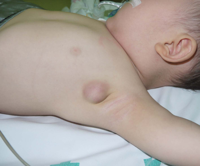 (
Image Source
)
Questions
- State the clinical diagnosis and distinguish between its two recognized clinical stages/forms.
- List three technical or vaccine-related risk factors contributing to the development of this Adverse Event Following Immunization (AEFI).
- Outline the evidence-based clinical management checklist for the non-suppurative versus suppurative forms of this condition.
- Explain why routine systemic anti-tubercular therapy (ATT) and incision and drainage (I&D) are contraindicated in this patient.
Answer
- Diagnosis & Classification:
- Diagnosis: BCG Lymphadenitis.
- Clinical Forms:
- Non-suppurative (simple/catarrhal) BCG lymphadenitis: Firm, mobile, non-tender enlargement of ipsilateral regional lymph nodes (axillary, supraclavicular, or cervical) $\gt 1\text{ cm}$, without overlying erythema or fluctuation; typically resolves spontaneously over 2–6 months.
- Suppurative BCG lymphadenitis: Progression to central caseation necrosis, fluctuation, purplish skin discoloration, thinning of overlying skin, and eventual spontaneous perforation forming a chronically draining sinus tract.
- Technical & Vaccine Risk Factors:
- Subcutaneous injection instead of strict intradermal delivery (most frequent technical fault).
- Inadvertent administration of an excessive vaccine volume or dose (e.g., using an incorrect syringe or failing to use an auto-disable $0.05\text{ mL}$ BCG syringe).
- High reactogenicity of specific vaccine strains (e.g., Pasteur 1173 P2, Danish 1331) compared to lower-reactogenicity strains (e.g., Tokyo 172).
- Administration in early neonates or low-birth-weight infants with immature cell-mediated immunity.
- Management Checklist:
- For Non-suppurative Lymphadenitis:
- Watchful waiting and regular clinical follow-up every 4–6 weeks.
- Parental reassurance that the lesion is a benign local immune response and typically resolves spontaneously without sequelae within 3 to 9 months.
- No pharmacological or surgical intervention indicated.
- For Suppurative / Fluctuant Lymphadenitis:
- Needle aspiration using a wide-bore needle ($18\text{--}20\text{ Gauge}$) inserted through intact normal adjacent skin (z-track technique) to evacuate purulent material.
- Repeat needle aspiration after 1–2 weeks if fluid re-accumulates.
- Total surgical excision of the node (under general anesthesia) reserved strictly for: failed needle aspiration, persistent matted draining multi-locular nodes, or large chronic draining sinuses.
- For Non-suppurative Lymphadenitis:
- Rationale Against Systemic ATT and Incision & Drainage:
- Why Systemic ATT is Contraindicated:
- Mycobacterium bovis BCG is inherently resistant to Pyrazinamide.
- Randomized trials demonstrate that oral anti-tubercular agents (such as Isoniazid, Rifampicin, or Erythromycin) do not shorten the duration of lymphadenopathy, prevent suppuration, or hasten resolution due to poor penetration into encapsulated necrotic lymph nodes.
- Why Incision & Drainage (I&D) is Contraindicated:
- Direct incision disrupts subcutaneous architecture and leads to chronic, non-healing sinus tracts, persistent purulent discharge, delayed secondary intention healing, and secondary pyogenic bacterial infection.
- Why Systemic ATT is Contraindicated:
RES010 - Neonatal Retroviral Exposure And Vaccination
Scenario
A term male neonate weighing 3.2 kg is delivered via elective cesarean section at 38 weeks of gestation to a 26-year-old primigravida. The mother was diagnosed with HIV-1 infection at 32 weeks of gestation and was immediately commenced on triple-drug Antiretroviral Therapy (Tenofovir Disoproxil Fumarate + Lamivudine + Dolutegravir). Her viral load at delivery is 850 copies/mL. The neonate is vigorous, pink, has a normal APGAR score of 9 at 5 minutes, and exhibits no dysmorphic features or organomegaly. The pediatric resident on duty asks for guidance regarding the administration of the live-attenuated BCG vaccine at birth.
Questions
- State the current WHO and National AIDS Control Organization (NACO) guidelines regarding the administration of the BCG vaccine to an asymptomatic HIV-exposed newborn at birth.
- Identify the specific clinical conditions and immunological cut-offs where the BCG vaccine is strictly contraindicated in HIV-exposed or infected children.
- Name the severe disseminated complication associated with administering BCG to an infant with severe HIV-related immunodeficiency.
- Formulate the therapeutic management plan if an infant inadvertently receives BCG vaccine and subsequently develops disseminated BCG disease (BCG-osis).
Answer
Guidelines on BCG at Birth in Asymptomatic HIV-Exposed Newborns:
- WHO & NACO Guidelines: In settings with high endemicity of tuberculosis (such as India), the benefits of BCG vaccination outweigh the potential risks. Therefore, BCG should be administered at birth to all asymptomatic HIV-exposed infants, regardless of maternal viral load or treatment status.
- Exception: Vaccination should be deferred only if the infant presents with clinical signs, symptoms, or severe illness suggestive of symptomatic HIV infection at birth.
Contraindications and Immunological Cut-Offs:
- Confirmed HIV infection in infants or children with documented severe immunosuppression:
- Children $<5\text{ years}$ of age: CD4+ T-cell percentage $<25\%$ (or CD4 absolute count $<750\text{ cells/}\mu\text{L}$ in $<12\text{ months}$; $<500\text{ cells/}\mu\text{L}$ in $1\text{–}5\text{ years}$).
- Children $\ge 5\text{ years}$ of age: CD4 absolute count $<200\text{ cells/}\mu\text{L}$ (or CD4 percentage $<15\%$).
- Any symptomatic HIV infection classified as WHO Clinical Stage 3 or Stage 4 disease.
- Primary immunodeficiency disorders (e.g., Severe Combined Immunodeficiency [SCID], Chronic Granulomatous Disease [CGD], Complete DiGeorge Syndrome, IFN-$\gamma$/IL-12 pathway defects).
- Confirmed HIV infection in infants or children with documented severe immunosuppression:
Disseminated Complication:
- Disseminated BCG Disease (Systemic BCG-osis): Life-threatening, systemic mycobacteremia characterized by Mycobacterium bovis BCG dissemination to two or more anatomical sites outside the regional drainage area (e.g., lungs, liver, spleen, bone marrow, bone/joints, CNS), with a mortality rate approaching $50\text{–}70\%$ in severely immunocompromised infants.
Therapeutic Management of Disseminated BCG-osis:
- Antimycobacterial Chemotherapy:
- Note: Mycobacterium bovis (including the BCG Moreau/Danish/Pasteur strains) has intrinsic resistance to Pyrazinamide (PZA) due to a point mutation in the pncA gene. Therefore, PZA must NOT be used.
- Multi-drug Regimen (minimum 4 drugs):
- High-dose Isoniazid ($15\text{–}20\text{ mg/kg/day}$, max $300\text{ mg}$) + Pyridoxine ($10\text{–}25\text{ mg/day}$).
- Rifampicin ($15\text{–}20\text{ mg/kg/day}$, max $600\text{ mg}$).
- Ethambutol ($20\text{–}25\text{ mg/kg/day}$).
- Plus a second-line bactericidal agent: Fluoroquinolone (Levofloxacin $15\text{–}20\text{ mg/kg/day}$ or Moxifloxacin $10\text{ mg/kg/day}$) or injectable Aminoglycoside (Amikacin $15\text{ mg/kg/day}$).
- Duration: Minimum 9 to 12 months of therapy (or at least 6 months following complete clinical and immunological recovery).
- Immunological & Antiretroviral Management:
- Prompt initiation or continuation of Antiretroviral Therapy (ART) to restore CD4 cell counts, with vigilance for paradoxical Immune Reconstitution Inflammatory Syndrome (IRIS).
- Antimycobacterial Chemotherapy:
RES011 - Neonatal Cyclical Cyanosis at Birth
Scenario
A term female neonate (birth weight 3,150 g, born at 39 weeks of gestation via uncomplicated vaginal delivery) presents in the labor room with severe respiratory distress, marked sternal retractions, and peripheral and central cyanosis immediately after birth. Direct laryngoscopic examination reveals normal vocal cord anatomy and an unobstructed glottic inlet. When the infant cries vigorously, the cyanosis promptly resolves and she turns completely pink, but severe distress recur whenever she closes her mouth or attempts to breastfeed. A bedside attempt to advance a lubricated 6 French suction catheter through both anterior nares fails to pass beyond 1.5 cm bilaterally.
Questions
- What is the most likely clinical diagnosis?
- What is the immediate bedside emergency intervention to secure the infant's airway?
- What is the gold-standard diagnostic imaging investigation to confirm the lesion and aid surgical planning?
- Name the well-recognized syndromic association linked with this condition, detail its diagnostic acronym, and identify the underlying causative gene.
Answer
- Primary Diagnosis:
- Bilateral choanal atresia (bony, membranous, or mixed bony-membranous).
- Immediate Bedside Emergency Airway Management:
- Immediate insertion of an oral Guedel airway or an adapted McGovern nipple (a modified feeding nipple with an enlarged distal opening secured in the oral cavity via ties) to maintain an oral airway bypass.
- Supplemental humidified oxygen via an open mouth route; endotracheal intubation if oral airway fails to alleviate severe asphyxia.
- Definitive Diagnostic Imaging Modality:
- Non-contrast high-resolution computed tomography (HRCT) of the paranasal sinuses and skull base (axial and coronal views with thin 1-mm slices), performed after suctioning nasal secretions and instilling topical nasal decongestants.
- Syndromic Association, Components, and Genetics:
- Syndrome: CHARGE syndrome.
- Acronym Components:
- C: Coloboma (most commonly chorioretinal or iridal).
- H: Heart defects (e.g., tetralogy of Fallot, ventricular septal defect, PDA).
- A: Atresia of choanae (unilateral or bilateral).
- R: Retardation of growth and development / CNS anomalies.
- G: Genital hypoplasia (micropenis, cryptorchidism, hypogonadotropic hypogonadism).
- E: Ear abnormalities / deafness (sensorineural or conductive hearing loss, dysplastic pinnae).
- Causative Gene: Heterozygous mutation or deletion of the CHD7 gene (chromodomain helicase DNA-binding protein 7) on chromosome 8q12.2.
More Details
RES012 - Pediatric Expiratory Wheezing Spirometry Evaluation
Scenario
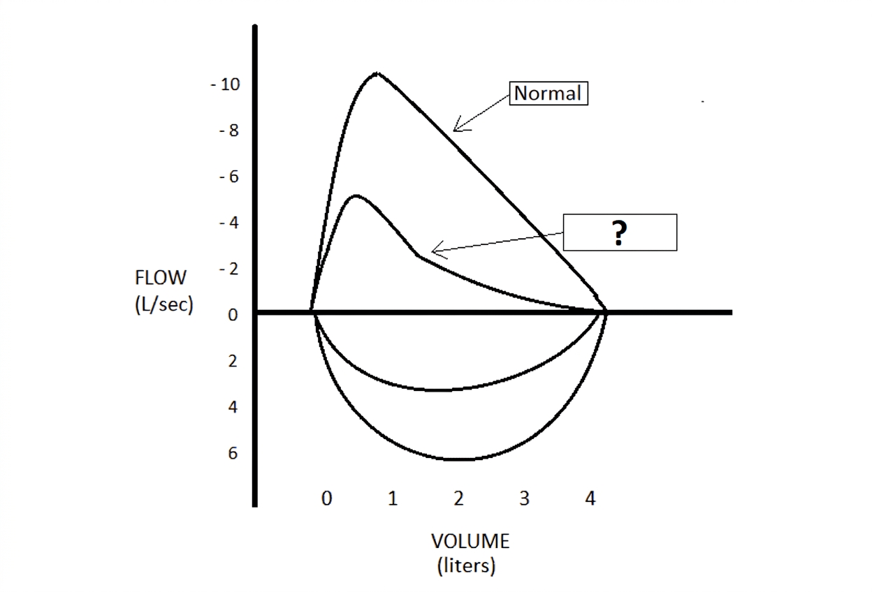
A 9-year-old boy (weight 28 kg) is brought to the pediatric pulmonary clinic with an 8-month history of recurrent nighttime dry coughing, breathlessness, and episodic chest tightness that worsens during physical education class and viral upper respiratory infections. On physical examination, respiratory rate is 22 breaths/min, oxygen saturation is 97% on room air, and prolonged expiration with bilateral end-expiratory polyphonic wheezing is auscultated over both lung fields. Spirometry is performed before and 15 minutes after the administration of 400 mcg of inhaled salbutamol delivered via a metered-dose inhaler with a spacer.
Questions
- Define the characteristic spirometric abnormalities observed during active, untreated disease in this child.
- What are the objective quantitative criteria defining significant bronchodilator reversibility and a positive exercise challenge test according to GINA/ATS guidelines?
- State the mathematical formula used to calculate Peak Expiratory Flow Rate (PEFR) diurnal variability and specify the cutoff threshold supporting this diagnosis.
- According to GINA (Global Initiative for Asthma) pediatric guidelines (ages 6–11 years), outline the initial step 2 controller therapy regimen and appropriate rescue medication.
Answer
- Baseline Spirometric Abnormalities:
- Reduced Forced Expiratory Volume in 1 second ($\text{FEV}_1$): $<80\%$ of predicted.
- Reduced $\text{FEV}_1/\text{FVC}$ ratio: $<0.80$ (or $<80\%$, or below the lower limit of normal [LLN]), indicating an obstructive ventilatory defect.
- Reduced mid-expiratory flow rates: $\text{FEF}_{25\text{–}75\%} < 65\%$ of predicted.
- Diagnostic Criteria for Reversibility and Exercise Challenge:
- Bronchodilator Reversibility: An increase in $\text{FEV}_1$ of $\ge 12\%$ from baseline (and/or $\ge 200\text{ mL}$ absolute increase in older children/adolescents) 10–15 minutes after inhaling 200–400 mcg of salbutamol.
- Exercise Challenge Test: A post-exercise fall in $\text{FEV}_1$ of $>10\%$ (or $>15\%$) from baseline measured at 5, 10, 15, and 30 minutes post-standardized exercise protocol.
- PEFR Diurnal Variability:
$$\begin{aligned}
\text{Daily Diurnal PEF Variability (\%)} &= \frac{\text{PEF}_{\text{highest}} - \text{PEF}_{\text{lowest}}}{\text{Mean}(\text{PEF}_{\text{highest}}, \text{PEF}_{\text{lowest}})} \times 100 \\
&\quad \text{or} \quad \frac{\text{PEF}_{\text{highest}} - \text{PEF}_{\text{lowest}}}{\text{PEF}_{\text{highest}}} \times 100
\end{aligned}
$$
- Diagnostic Cutoff: Daily diurnal PEFR variability $>13\%$ in children (calculated over 1–2 weeks of twice-daily monitoring).
- Step 2 Maintenance and Reliever Therapy (GINA Ages 6–11 Years):
- Preferred Controller: Low-dose Inhaled Corticosteroid (ICS) daily maintenance:
- Inhaled Fluticasone propionate: 50 mcg twice daily (100 mcg/day total), OR
- Inhaled Budesonide: 100–200 mcg twice daily via MDI with valved holding chamber.
- Alternative Controller: Leukotriene receptor antagonist (Montelukast 5 mg orally once daily at bedtime).
- Reliever Therapy: Inhaled Short-Acting $\beta_2$-Agonist (SABA; Salbutamol 100–200 mcg [1–2 puffs] via MDI with spacer) as needed for symptom relief.
- Preferred Controller: Low-dose Inhaled Corticosteroid (ICS) daily maintenance:
RES013 - Chronic Productive Cough Imaging Assessment
Scenario
An 8-year-old girl is evaluated in the pediatric outpatient department for a chronic, productive cough persisting for 7 months. Her parents report expectoration of copious, three-layered, purulent, foul-smelling sputum, most prominent upon waking in the morning, with two recent episodes of blood-streaked phlegm (mild hemoptysis). She has been treated with five courses of oral antibiotics over the past half-year for recurrent lower respiratory infections. Physical examination reveals grade 3 digital clubbing, mild pectus excavatum, coarse bubbling mid-inspiratory and late-inspiratory crackles over the left lower lung zone, and persistent expiratory wheezing. Non-contrast High-Resolution Computed Tomography (HRCT) of the chest is performed.
Questions
- Enumerate five key thoracic HRCT signs diagnostic of this clinical entity.
- Outline the Reid pathological classification of this structural airway disorder.
- Formulate a structured etiologic diagnostic workup consisting of four targeted investigations to determine the underlying cause in this child.
- Detail the primary components of long-term medical management, including airway clearance techniques and antimicrobial therapy.
Answer
- Diagnostic HRCT Features:
- Increased Bronchoarterial Ratio: Internal diameter of the bronchus exceeding the diameter of its accompanying pulmonary artery (bronchoarterial ratio $>0.80\text{–}1.0$), producing the "signet ring sign".
- Lack of Bronchial Tapering: Parallel bronchial walls failing to taper progressively toward the periphery for $>2\text{ cm}$ distal to a bifurcation ("tram-track" sign on longitudinal cuts).
- Peripheral Bronchial Visualization: Identification of non-tapering bronchi within $1\text{ cm}$ of the costal pleural surface or abutting the mediastinal pleura.
- Bronchial Wall Thickening: Prominent peribronchial interstitial cuffing.
- Mucus Impaction: Tubular branching or rounded fluid/mucus opacities creating the "finger-in-glove" sign or "tree-in-bud" nodularity reflecting associated bronchiolitis.
- Reid Pathological Classification:
- Cylindrical (Tubular) Bronchiectasis: Uniformly dilated, smoothly contoured airways without normal tapering; bronchial divisions remain normal.
- Varicose Bronchiectasis: Irregular, beaded bronchial dilatations alternating with constricted areas, resembling varicose veins.
- Saccular (Cystic) Bronchiectasis: Severe, progressive ballooning of bronchi ending in blind, fluid-filled or air-filled fibrous sacs clustered like a bunch of grapes; extensive loss of distal bronchial subdivisions.
- Structured Pediatric Etiologic Workup:
- Cystic Fibrosis Evaluation: Quantitative pilocarpine iontophoresis sweat chloride testing (normal $<30\text{ mmol/L}$, intermediate $30\text{–}59\text{ mmol/L}$, positive $\ge 60\text{ mmol/L}$) and CFTR genetic mutation analysis.
- Primary Ciliary Dyskinesia (PCD): Nasal nitric oxide (nNO) measurement (markedly reduced, often $<77\text{ nL/min}$), high-speed video microscopy analysis (HSVA) of ciliary beat frequency/pattern, and transmission electron microscopy or ciliary genetic panel.
- Immunodeficiency Screening: Serum quantitative immunoglobulin levels (IgG, IgA, IgM, IgE), IgG subclass levels, and post-vaccination specific antibody titers against tetanus toxoid and Streptococcus pneumoniae.
- Aspiration / Structural Assessment: Fiberoptic bronchoscopy with bronchoalveolar lavage (bacterial, fungal, mycobacterial cultures, lipid-laden macrophage index) and barium videofluoroscopic swallow study.
- Long-Term Medical Management Plan:
- Airway Clearance Therapy (ACT): Twice-daily chest physiotherapy using positive expiratory pressure (PEP) devices, oscillating PEP (e.g., Flutter valve, Acapella), or high-frequency chest wall oscillation combined with active cycle of breathing techniques (ACBT).
- Adjunctive Inhalation Therapy: Inhaled hypertonic saline (3% or 7% via nebulizer twice daily) before physiotherapy to rehydrate secretions; short-acting bronchodilator pre-treatment to mitigate bronchospasm.
- Antimicrobial Strategy:
- Acute Exacerbations: Sputum culture-directed high-dose oral or intravenous antibiotics for 14 days (e.g., Amoxicillin-clavulanate, or Ciprofloxacin / Ceftazidime if Pseudomonas aeruginosa isolated).
- Long-Term Prophylaxis: For frequent exacerbators ($\ge 3$ per year), maintenance oral Azithromycin ($10\text{ mg/kg}$ thrice weekly or $5\text{ mg/kg/day}$ once daily) for its immunomodulatory and anti-inflammatory properties.
RES014 - Flow Volume Loop Reversibility Analysis
Scenario
An 11-year-old boy with chronic exercise limitation and nocturnal coughing attends the pulmonary physiology laboratory for formal evaluation. Baseline maximal expiratory-inspiratory flow-volume loops are generated. He is then administered 4 puffs (400 mcg) of inhaled salbutamol via an MDI with an antistatic spacer chamber. Spirometric measurements are repeated after a 15-minute rest period.
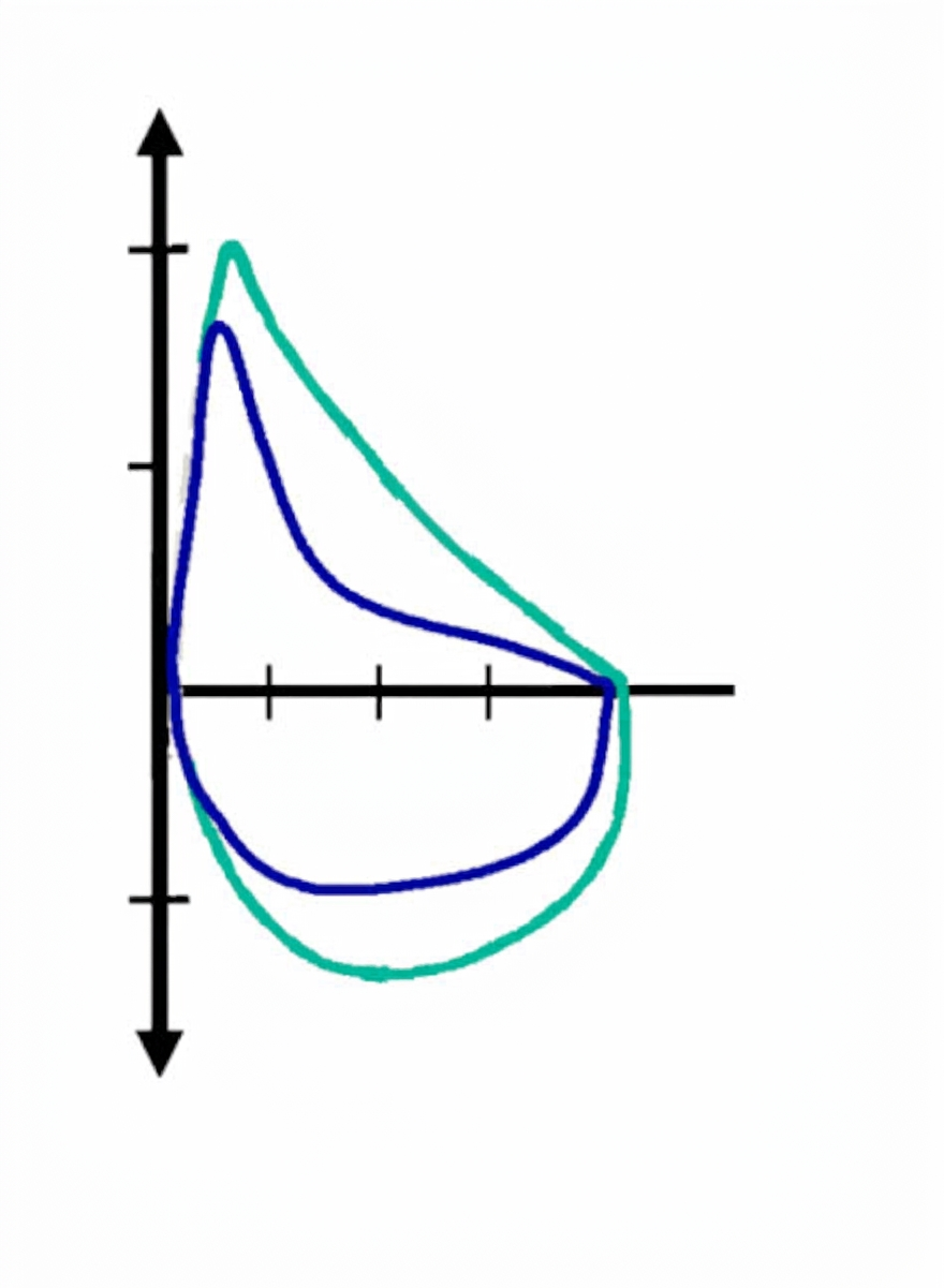
The laboratory computer produces the following spirometric values:
- Baseline (Pre-bronchodilator):
- $\text{FEV}_1 = 1.20\text{ L}$ ($48\%$ of predicted)
- $\text{FVC} = 2.50\text{ L}$ ($85\%$ of predicted)
- $\text{FEV}_1/\text{FVC} = 0.48$ ($48\%$)
- Post-bronchodilator:
- $\text{FEV}_1 = 1.56\text{ L}$ ($62\%$ of predicted)
- $\text{FVC} = 2.65\text{ L}$ ($90\%$ of predicted)
- $\text{FEV}_1/\text{FVC} = 0.59$ ($59\%$)
Questions
- Describe the characteristic morphological abnormality displayed by the expiratory limb of the baseline (in blue) flow-volume loop.
- Calculate the absolute change (in mL) and the percentage bronchodilator reversibility of $\text{FEV}_1$.
- Based on standardized ATS/ERS international pediatric guidelines, does this child demonstrate statistically and clinically significant bronchodilator reversibility? State the defining cutoff criteria.
- Explain the physical mechanism underlying the concave or "scooped-out" contour of the descending expiratory limb during forced expiration.
Answer
- Baseline Loop Morphology:
- Marked upward concavity ("scooped-out" appearance) of the descending portion of the maximal expiratory flow-volume curve, with a decreased peak expiratory flow (PEF) and disproportionate reduction in instantaneous mid-to-late flows ($\text{FEF}_{50\%}$ and $\text{FEF}_{75\%}$).
- Calculation of Bronchodilator Reversibility: $$\begin{aligned} \text{Absolute Change in FEV}_1 &= \text{Post-BD FEV}_1 - \text{Pre-BD FEV}_1 \\ &= 1.56\text{ L} - 1.20\text{ L} \\ &= \mathbf{0.36\text{ L}} \quad (\mathbf{360\text{ mL}}) \end{aligned} $$ $$\begin{aligned} \text{Percentage Change in FEV}_1 &= \frac{\text{Post-BD FEV}_1 - \text{Pre-BD FEV}_1}{\text{Pre-BD FEV}_1} \times 100 \\ &= \frac{1.56 - 1.20}{1.20} \times 100 \\ &= \frac{0.36}{1.20} \times 100 \\ &= \mathbf{30.0\%} \end{aligned} $$
- Interpretation Against Standardized Criteria:
- Yes, the test demonstrates highly significant bronchodilator reversibility.
- Standard Cutoff Criteria: An increase in $\text{FEV}_1 \ge 12\%$ relative to the baseline value AND an absolute increase $\ge 200\text{ mL}$ (both criteria are exceeded: $30.0\%$ and $360\text{ mL}$).
- Biomechanical Mechanism of Concavity:
- During forced expiration, positive intrathoracic pleural pressure compresses small intrathoracic, non-cartilaginous airways downstream of the equal pressure point (dynamic airway compression).
- In obstructive airway disease, intrinsic luminal narrowing, mucosal edema, and loss of elastic parenchymal radial tethering cause premature airway closure and dynamic collapse, leading to an abrupt, precipitous drop in airflow at lower lung volumes that manifests visually as an expiratory scoop.
RES015 - Pediatric Persistent Mediastinal Mass Evaluation
Scenario
A 5-year-old girl is evaluated for recurrent episodes of right lower lobe wheezing and cough refractory to multiple courses of inhaled bronchodilators and antibiotics. She has no history of choking, dysphagia, or failure to thrive. Physical examination reveals decreased breath sounds and transmission of localized monophonic wheeze over the right infrascapular area. A chest radiograph demonstrates a smooth, sharply demarcated round opacity projected over the right retrocardiac and subcarinal mediastinum. Magnetic resonance imaging (MRI) of the thorax confirms a homogeneous, sharply marginated, unilocular $4.5 \times 3.8\text{ cm}$ cystic mass located in the middle mediastinum immediately subcarinal to and compressing the right mainstem bronchus; it is hypointense on T1-weighted images, hyperintense on T2-weighted images, and exhibits no contrast enhancement or communication with the esophageal lumen or spinal canal.
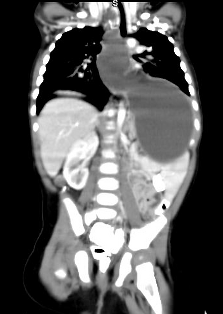 (
Image Source
)
Questions
- What is the most definitive clinical diagnosis?
- Detail the embryological origin of this malformation and state the pathognomonic histopathological epithelial lining and wall constituents that establish the diagnosis.
- List four distinct differential diagnoses for congenital or neoplastic cystic mediastinal masses occurring in this compartment in a young child.
- What is the definitive management of choice, and what two major long-term complications mandate intervention even in asymptomatic children?
Answer
- Primary Diagnosis:
- Mediastinal Bronchogenic Cyst (Foregut Duplication Cyst).
- Embryological Origin and Histopathological Hallmarks:
- Embryology: Abnormal budding or branching of the ventral diverticulum of the primitive embryonic foregut during the 3rd to 7th weeks of gestation.
- Histopathology:
- Cyst lining composed of ciliated pseudostratified columnar respiratory epithelium.
- Cyst wall contains elements of the bronchial wall: hyaline cartilage plates, smooth muscle bundles, fibrous connective tissue, and mucus-secreting seromucinous bronchial glands (the presence of cartilage plates definitively differentiates it from esophageal duplication cysts).
- Differential Diagnosis of Middle/Posterior Mediastinal Cystic Masses:
- Esophageal duplication cyst (enteric cyst; lined by gastrointestinal mucosa with a dual smooth muscle coat, lacking cartilage).
- Neurenteric cyst (associated with vertebral segmentation anomalies like hemivertebrae).
- Mediastinal lymphatic malformation (cystic hygroma / lymphangioma).
- Pericardial / pleuropericardial cyst.
- Definitive Management and Long-Term Complications:
- Management: Complete surgical excision (via video-assisted thoracoscopic surgery [VATS] or open posterolateral thoracotomy) with preservation of adjacent tracheobronchial and esophageal structures.
- Complications Mandating Excision:
- Secondary infection and abscess formation: Intracystic infection with potential rupture into the pleural space (empyema) or bronchial tree (bronchopleural/cystobronchial fistula).
- Progressive tracheobronchial compression: Severe extrinsic tracheomalacia, atelectasis, air-trapping, or secondary bronchiectasis.
- Malignant transformation: Rare risk of malignant degeneration in adulthood (e.g., adenocarcinoma, rhabdomyosarcoma).
RES016 - Childhood Nocturnal Sleep Disturbance
Scenario
An 8-year-old boy is brought to the pediatric clinic by his parents due to loud, persistent snoring for the past 9 months. The mother notes frequent pauses in his breathing followed by gasping and restlessness during sleep. He is difficult to awaken in the morning, exhibits daytime somnolence, and has recently shown academic decline with hyperactivity. Physical examination reveals a body mass index (BMI) of 18.2 kg/m² (75th percentile), mouth breathing, tonsillar hypertrophy grade 3+, and high-arched palate. An overnight sleep study is conducted.
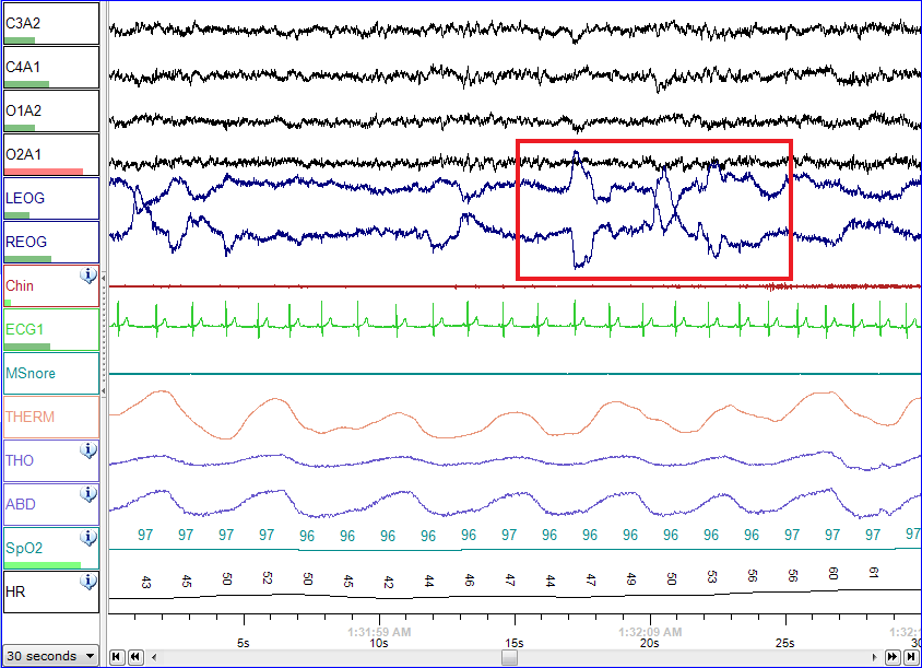 (
Image Source
)
Questions
- Identify the diagnostic investigation demonstrated in this trace.
- Enlist six physiological parameters monitored simultaneously during this assessment.
- State the diagnostic criteria and severity grading for this condition in children based on the Apnea-Hypopnea Index (AHI).
- Outline the definitive first-line intervention and two non-surgical medical treatment options for mild or persistent disease.
Answer
- Diagnostic Investigation: Overnight Polysomnography (Level 1 Sleep Study).
- Physiological Parameters Monitored:
- Electroencephalography (EEG: frontal, central, occipital leads for sleep staging)
- Electrooculography (EOG: bilateral eye movements)
- Electromyography (EMG: submental chin tone and bilateral anterior tibialis leads)
- Electrocardiography (ECG / continuous heart rate)
- Respiratory airflow (nasal pressure transducer and oronasal thermistor)
- Respiratory effort (thoracoabdominal dual-band inductance plethysmography)
- Oxygen saturation ($SpO_2$ via continuous pulse oximeter) and carbon dioxide monitoring (end-tidal $EtCO_2$ or transcutaneous $TcCO_2$)
- Diagnostic Criteria and Pediatric Severity Grading:
- Diagnostic threshold: Obstructive Apnea-Hypopnea Index (oAHI) $\ge 1$ event per hour of total sleep time.
- Severity categorization:
- Mild: oAHI $1.0$ to $< 5.0$ events/hour
- Moderate: oAHI $5.0$ to $< 10.0$ events/hour
- Severe: oAHI $\ge 10.0$ events/hour (or minimum nocturnal $SpO_2$ nadir $< 80\%$)
- Management:
- First-line surgical treatment: Adenotonsillectomy (treatment of choice in adenotonsillar hypertrophy).
- Medical options:
- Intranasal corticosteroids (Fluticasone furoate or Mometasone spray) combined with oral Leukotriene Receptor Antagonists (Montelukast 5 mg once daily) for 6–12 weeks in mild disease or post-surgical residual disease.
- Continuous Positive Airway Pressure (CPAP) or Bi-level Positive Airway Pressure (BiPAP) if surgery is contraindicated, craniofacial abnormalities are present, or severe OSA persists post-adenotonsillectomy.
RES017 - Neonatal Oromandibular Midline Defect
Scenario
A male infant born at 38 weeks of gestation (birth weight 3.1 kg) to a 26-year-old primigravida is assessed in the neonatal nursery. Delivery room inspection reveals an orofacial cleft defect. Antenatal history reveals irregular attendance; a routine 20-week anomaly scan was performed and reportedly called unremarkable. The baby exhibits normal vigor and respiratory effort but exhibits uncoordinated sucking.
Questions
- Determine the validity (True or False) of the following clinical statements regarding orofacial clefts:
- (a) Isolated cleft palate without cleft lip is routinely and reliably detected on standard 20-week screening ultrasound.
- (b) Periconceptional maternal folic acid supplementation reduces the incidence of non-syndromic cleft lip and palate.
- (c) Neonates with an isolated cleft lip generally experience more severe mechanical feeding compromise than neonates with an isolated cleft palate.
- Name two genetic syndromes or developmental sequences characteristically associated with cleft palate.
- State the surgical criteria (Wilhelmsen and Musgrave "Rule of 10s") for repairing the primary lip defect, and give the optimal age window for palatoplasty.
- Enlist two middle ear complications that require routine audiologic surveillance in these children, explaining the underlying anatomical mechanism.
Answer
- True/False Statements:
- (a) False: The fetal secondary palate is obscured by facial bones and acoustic shadowing; prenatal 2D ultrasound detects $< 15–20\%$ of isolated cleft palates (whereas cleft lip is easily identified on coronal facial views).
- (b) True: Daily periconceptional supplementation with folic acid (400 mcg to 4 mg) significantly lowers the risk of non-syndromic orofacial clefting.
- (c) False: Isolated cleft lip preserves the intraoral seal and hard palate plate against which the nipple is compressed, allowing normal suction. Cleft palate prevents negative pressure generation, resulting in severe sucking impairment and nasal regurgitation.
- Associated Syndromes/Sequences:
- Pierre Robin sequence (micrognathia, glossoptosis, U-shaped cleft palate)
- 22q11.2 Deletion syndrome (Velocardiofacial / DiGeorge syndrome)
- Stickler syndrome
- Van der Woude syndrome
- Surgical Timing:
- Wilhelmsen and Musgrave "Rule of 10s" for Cleft Lip Repair:
- Age $\ge 10$ weeks
- Weight $\ge 10$ pounds ($\approx 4.5\text{ kg}$)
- Hemoglobin $\ge 10\text{ g/dL}$
- White blood cell (WBC) count $< 10,000/\text{mm}^3$
- Palatoplasty timing: 9 to 12 months of age (completed prior to development of compensatory articulation patterns, while mitigating midface hypoplasia).
- Wilhelmsen and Musgrave "Rule of 10s" for Cleft Lip Repair:
- Middle Ear Complications and Mechanism:
- Complications: Recurrent acute otitis media, Otitis Media with Effusion (OME / "glue ear"), and Conductive Hearing Loss.
- Mechanism: Abnormal insertion and hypoplasia of the tensor veli palatini and levator veli palatini muscles into the posterior border of the hard palate instead of forming a midline aponeurosis, leading to Eustachian tube dysfunction and impaired middle ear ventilation.
RES018 - Pediatric Chronic Meningitic Presentation
Scenario
A 4-year-old girl is admitted with a 3-week history of low-grade fever, worsening irritability, anorexia, and persistent dry cough. Her weight is 11.2 kg ($< 3\text{rd}$ percentile for age). The child's paternal grandmother died of pulmonary tuberculosis one month prior to admission. On hospital day 4, the child develops lethargy, terminal neck stiffness, right complete ptosis with pupillary dilatation, and left hemiparesis. Urgent neuroimaging and lumbar puncture are organized.
Questions
- Enlist four cardinal findings visualized on contrast-enhanced CT (CECT) or MRI brain characteristic of tuberculous meningitis (TBM).
- Describe the classic cerebrospinal fluid (CSF) cytochemical profile in central nervous system tuberculosis.
- State the British Medical Research Council (BMRC) clinical staging system for TBM, and assign the clinical stage for this patient.
- Detail the medical management plan: specify the four-drug anti-tubercular therapy (ATT) regimen with pediatric weight-based dosing, total duration, and systemic corticosteroid dosing protocol.
Answer
- Cardinal Neuroimaging Findings:
- Marked, thick basal meningeal enhancement (in suprasellar cistern, perimesencephalic cistern, and Sylvian fissures)
- Hydrocephalus (communicating due to arachnoid exudates blocking arachnoid villi > non-communicating/obstructive)
- Vasculitic cerebral infarcts (tuberculous endarteritis obliterans classically affecting the medial striate and lenticulostriate branches in the tuberculous "tubercular vascular zone" – basal ganglia and internal capsule)
- Tuberculomas (single or multiple ring-enhancing or nodular granulomatous parenchymal lesions)
- CSF Cytochemical Profile:
- Appearance: Clear or xanthochromic, often forming a "cobweb" or "spiderweb" fibrin clot/coagulum on refrigeration
- Cell count: Pleocytosis of $50–500\text{ cells/}\mu\text{L}$, predominantly lymphocytic/mononuclear (may have transient neutrophilic predominance in the initial 24–48 hours)
- Protein: Markedly elevated ($100–500\text{ mg/dL}$; can exceed $1000\text{ mg/dL}$ in spinal block)
- Glucose: Markedly reduced with a CSF-to-blood glucose ratio $< 0.30$ (absolute CSF glucose often $< 40\text{ mg/dL}$)
- Chloride: Reduced ($< 110\text{ mEq/L}$) due to systemic hypochloremia and SIADH
- BMRC Staging of TBM:
- Stage 1: Fully conscious, alert, non-specific symptoms (fever, headache, irritability), no neurological deficits, no meningeal signs.
- Stage 2: Conscious but irritable or lethargic, signs of meningism, with focal neurological deficits (cranial nerve palsies or hemiparesis), but no coma.
- Stage 3: Severely altered sensorium (stupor or coma), dense paraplegia/hemiplegia, or decerebrate/decorticate posturing.
- Patient Stage: BMRC Stage 2 (lethargy, cranial nerve III palsy, and left hemiparesis).
- Medical Management Protocol:
- Anti-Tubercular Therapy (Total 12 months):
- Intensive Phase (2 months - HRZE):
- Isoniazid (H): $10\text{ mg/kg/day}$ (range 7–15) PO
- Rifampicin (R): $15\text{ mg/kg/day}$ (range 10–20) PO
- Pyrazinamide (Z): $35\text{ mg/kg/day}$ (range 30–40) PO
- Ethambutol (E): $20\text{ mg/kg/day}$ (range 15–25) PO
- Continuation Phase (10 months - HRE):
- Isoniazid + Rifampicin + Ethambutol daily for 10 months.
- Pyridoxine supplementation: $10–25\text{ mg/day}$ to prevent INH-induced peripheral neuropathy.
- Intensive Phase (2 months - HRZE):
- Systemic Corticosteroids:
- Dexamethasone $0.4\text{ mg/kg/day}$ IV (or Prednisolone $2–4\text{ mg/kg/day}$ PO) for 4 weeks.
- Taper gradually over the subsequent 4 weeks (total corticosteroid duration: 8 weeks).
- Anti-Tubercular Therapy (Total 12 months):
RES019 - Pediatric Hearing Evaluation Profile
Scenario
A 3-year-old girl is brought by her mother because of speech delay and suspected hearing difficulty. She frequently speaks loudly, fails to respond to conversational verbal requests unless directly facing the speaker, and increases the volume on audio devices. She has had three episodes of upper respiratory tract infections in the preceding 6 months. Otoscopy reveals dull, retracted bilateral tympanic membranes with prominent lateral malleolar processes and fluid levels. Pure tone audiometry is obtained.
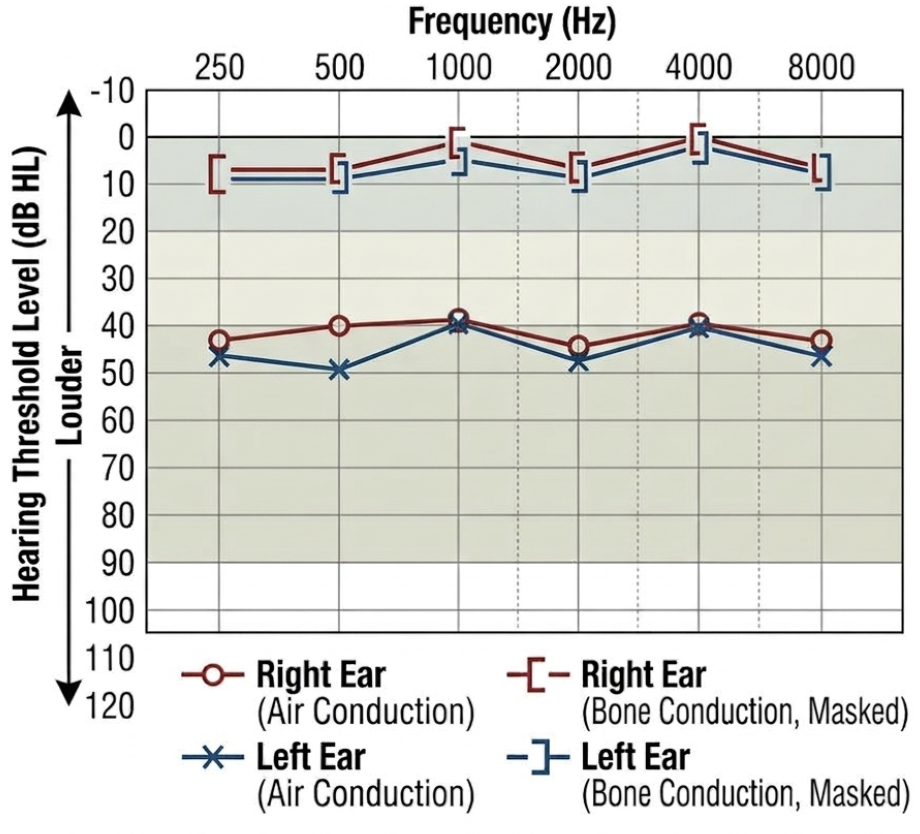
Questions
- State the audiometric interpretation and type of hearing impairment demonstrated in the exhibit.
- Define the exact audiometric threshold criteria (in decibels) that confirm this pattern over sensorineural impairment.
- Enlist four etiologies causing this type of auditory deficit in preschool children.
- Detail the non-surgical observation strategy and specify the definitive surgical intervention with its precise clinical indications.
Answer
- Audiometric Interpretation: Bilateral Conductive Hearing Loss (CHL).
- Audiometric Criteria:
- Bone conduction thresholds are within normal limits ($\le 20\text{ dB HL}$).
- Air conduction thresholds are elevated ($> 20\text{ dB HL}$).
- Presence of an Air-Bone Gap (ABG) $\ge 15–20\text{ dB}$ across speech frequencies ($500\text{ Hz}$, $1000\text{ Hz}$, $2000\text{ Hz}$, $4000\text{ Hz}$).
- Etiologies in Preschool Children:
- Otitis Media with Effusion (OME / serous otitis media / "glue ear")
- Impacted cerumen (earwax)
- Acute otitis media (AOM) or Chronic suppurative otitis media (CSOM)
- Foreign body impaction in the external auditory canal
- Tympanic membrane perforation or ossicular chain discontinuity/fixation
- Management Strategy:
- Observation / Medical Approach:
- Watchful waiting for 3 months from initial diagnosis, as up to $75–90\%$ of acute OME episodes resolve spontaneously.
- Treat predisposing allergic rhinitis if present.
- Antihistamines, oral/topical decongestants, systemic steroids, and routine prophylactic antibiotics are not recommended.
- Surgical Intervention:
- Procedure: Bilateral Myringotomy with Tympanostomy Tube (Grommet) insertion $\pm$ Adenoidectomy (if concurrent adenoid hypertrophy or recurrent rhinosinusitis).
- Indications:
- Bilateral OME persisting $\ge 3$ months with documented bilateral hearing impairment (pure tone threshold $> 20–25\text{ dB}$).
- Documented speech and language delay or academic performance deficits attributable to OME.
- Structural changes to the tympanic membrane (e.g., retraction pocket, atelectasis, adhesive otitis, or ossicular erosion).
- Observation / Medical Approach:
RES020 - Neonatal Perinatal Mycobacterial Exposure
Scenario
A 10-day-old male neonate born at 38 weeks of gestation (birth weight 2.85 kg) is brought to the pediatric clinic. Three days post-partum, the mother was evaluated for chronic productive cough of 3 months' duration and evening fever; her sputum cartridge-based nucleic acid amplification test (CBNAAT / GeneXpert MTB/RIF) detected Mycobacterium tuberculosis (rifampicin-sensitive), and her chest radiograph revealed bilateral apical cavitary lesions. The infant is currently asymptomatic, afebrile, and breastfeeding well.
Questions
- Differentiate the primary anatomical sites of tuberculous complex formation in congenital TB acquired via the transplacental route versus the intrapartum aspiration route.
- State the four Modified Cantwell Criteria used to confirm a diagnosis of congenital tuberculosis.
- Outline the immediate clinical and laboratory diagnostic workup required in this asymptomatic neonate to exclude active tuberculosis disease.
- Describe the specific pharmacologic preventive strategy (drug name, weight-based dose, duration), breastfeeding guidance, and the exact protocol for BCG vaccination in this infant.
Answer
- Primary Focus by Route of Acquisition:
- Transplacental (Hematogenous via Umbilical Vein): Primary complex develops in the Liver (hepatic parenchymal caseating granulomas and porta hepatis lymphadenopathy); secondarily seeds lungs hematogenously via inferior vena cava and ductus venosus.
- Aspiration / Ingestion (Infected Amniotic Fluid or Genital Secretions): Primary complex develops in the Lungs (alveolar complex with mediastinal adenopathy) or Gastrointestinal Tract (gut mucosal focus with mesenteric lymphadenopathy).
- Modified Cantwell Criteria:
- Proven tuberculous lesions (Mycobacterium tuberculosis demonstrated by smear, culture, or NAAT) in the infant, PLUS at least one of the following:
- Onset of disease symptoms within the first week of life.
- Primary hepatic complex or caseating hepatic granulomas on liver biopsy or autopsy.
- Documentation of tuberculosis infection in the placenta or maternal genital tract.
- Complete exclusion of postnatal transmission by rigorous investigation of all infant contacts.
- Proven tuberculous lesions (Mycobacterium tuberculosis demonstrated by smear, culture, or NAAT) in the infant, PLUS at least one of the following:
- Diagnostic Workup to Exclude Active Disease:
- Gastric aspirate on 3 consecutive mornings for smear microscopy (AFB) and CBNAAT/GeneXpert (and liquid mycobacterial culture).
- Chest radiography (anteroposterior and lateral views) to inspect for miliary infiltrates, parenchymal consolidation, or hilar adenopathy.
- Abdominal ultrasonography (evaluating for hepatosplenomegaly, hepatic microgranulomas, and enlarged retroperitoneal/mesenteric lymph nodes).
- Cerebrospinal fluid (CSF) analysis (cell count, protein, sugar, CBNAAT) if signs of sepsis or meningitis emerge.
- Preventive Therapy and Immunization Protocol:
- Chemoprophylaxis (TB Preventive Therapy - TPT):
- If diagnostic evaluation excludes active tuberculosis (asymptomatic infant, normal chest radiograph, and negative gastric aspirates for AFB/CBNAAT), initiate Isoniazid (INH) Preventive Therapy immediately at $10\text{ mg/kg/day}$ orally once daily, along with Pyridoxine ($10\text{ mg/day}$), for a duration of 6 months.
- Breastfeeding Guidance:
- The mother should safely continue exclusive breastfeeding. Breast milk does not transmit M. tuberculosis.
- She must wear a well-fitted surgical mask, cover her mouth and nose when coughing, and maintain hand hygiene when handling or feeding the baby.
- First-line anti-TB drugs excreted in breast milk are in negligible, subtherapeutic amounts; they cause no toxicity to the infant but do not provide prophylaxis, necessitating dedicated infant INH therapy.
- BCG Vaccination Protocol:
- Defer BCG vaccination at birth because prophylactic Isoniazid will destroy the live attenuated M. bovis BCG bacilli and render the vaccine ineffective.
- At the completion of the 6-month INH course, perform a Tuberculin Skin Test (Mantoux test, 2 TU PPD RT-23):
- If Mantoux is negative ($<5\text{ mm}$ induration): Discontinue INH and administer BCG vaccine ($0.05\text{ mL}$ intradermally).
- If Mantoux is positive ($\ge 5\text{ mm}$ induration): Re-evaluate rigorously (repeat CXR, physical exam, gastric aspirates) to rule out active disease; if active TB is confirmed, initiate full 4-drug pediatric ATT; if active disease is ruled out, complete another 3 months of TPT (total 9 months), and omit BCG.
- Chemoprophylaxis (TB Preventive Therapy - TPT):
RES021 - Persistent Childhood Fever And Cough
Scenario
A 4-year-old child presents to the pediatric emergency department with a 3-week history of low-to-moderate grade intermittent fever and a progressively worsening dry cough over the past 2 weeks. The child's paternal grandmother died of sputum-positive pulmonary tuberculosis 1 month ago.
Physical examination reveals:
- Weight: 11.2 kg (<3rd percentile for age and sex, WHO growth charts)
- Heart rate: 118 beats/min, Respiratory rate: 38 breaths/min, $SpO_2$: 91% on ambient air
- General examination: Severe pallor, generalized non-tender discrete lymphadenopathy
- Respiratory system: Bronchial breath sounds and dullness to percussion over the right mid-to-lower zones; bilateral fine crackles on auscultation
A chest radiograph is performed as part of the primary diagnostic evaluation.
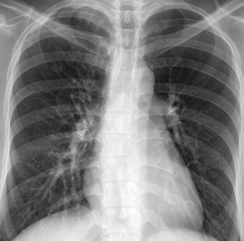 (
Image Source
)
Questions
- Based on the chest radiograph and clinical presentation, what are four established, evidence-based indications for adjunctive systemic corticosteroid therapy in pediatric tuberculosis?
- What are two relative or controversial clinical indications for adjunctive corticosteroid administration in pulmonary tuberculosis?
- What is the recommended systemic corticosteroid, dose, route, and tapering schedule for a child diagnosed with tuberculous meningitis (TBM)?
- Name two crucial baseline clinical parameters or adverse events that require vigilant monitoring during systemic corticosteroid therapy in a malnourished child receiving first-line anti-tubercular therapy (ATT).
Answer
- Established Indications for Adjunctive Corticosteroids in Pediatric Tuberculosis:
- Tuberculous meningitis (TBM): Reduces mortality, cerebral edema, hydrocephalus, and neurological sequelae.
- Tuberculous pericarditis: Decreases pericardial constriction, reduces the need for pericardiectomy, and accelerates resolution of large pericardial effusions.
- Miliary tuberculosis with acute respiratory failure / ARDS: Alleviates alveolar-capillary block, severe hypoxemia, and secondary hyperinflammatory lung injury.
- Primary adrenal tuberculosis (Addisonian crisis): Treats acute primary adrenocortical insufficiency and prevents circulatory collapse.
- Relative / Controversial Indications:
- Endobronchial tuberculosis with severe extrinsic airway compression: Tracheobronchial lymph node mass causing subtotal luminal obstruction, localized lobar hyperinflation, or atelectasis.
- Massive pleural effusion with respiratory compromise: Accelerates fluid reabsorption and shortens recovery, though it does not significantly prevent long-term pleural thickening.
- Paradoxical upgrade reactions / Immune Reconstitution Inflammatory Syndrome (IRIS): Significant clinical worsening following initial microbiological improvement on ATT.
- Corticosteroid Protocol for Tuberculous Meningitis:
- Drug & Dose: Oral Prednisolone at $2\text{ mg/kg/day}$ (or intravenous Dexamethasone $0.4\text{ mg/kg/day}$) administered in single or divided doses.
- Duration & Tapering: Full therapeutic dose for 4 weeks, followed by a gradual tapering over the subsequent 2 to 4 weeks (total duration: 6 to 8 weeks).
- Monitoring Parameters During Therapy:
- Blood pressure and serum glucose: Surveillance for steroid-induced secondary hypertension and hyperglycemia.
- Secondary bacterial/fungal infections & gastric ulceration: Vigilance for occult sepsis, clinical decompensation, and gastrointestinal hemorrhage.
RES022 - Pediatric Pulmonary Computed Tomography Interpretation
Scenario
A 7-year-old child is referred to the Pediatric Pulmonology clinic for evaluation of chronic productive cough lasting longer than 4 months, refractory to multiple empirical outpatient antibiotic courses. The team is reviewing high-resolution chest computed tomography (HRCT) distributions across various disease patterns to guide diagnostic testing.
Questions
- List four important pediatric differential diagnoses characterized by predominant bilateral upper lobe involvement on chest CT.
- What is the most common etiology when chest CT demonstrates predominant unilateral upper lobe consolidation, cavitation, or fibro-cavitary destruction in older pediatric patients?
- List four prominent etiologies characterized by predominant lower lobe distribution of bronchiectasis, consolidation, or interstitial changes in children.
- Name the classic CT sign seen when dilated bronchi with thickened walls accompany a matching branch of the pulmonary artery in cross-section.
Answer
- Differential Diagnoses for Bilateral Upper Lobe Involvement:
- Cystic fibrosis (CF)
- Allergic Bronchopulmonary Aspergillosis (ABPA)
- Pulmonary sarcoidosis
- Langerhans cell histiocytosis (LCH)
- Silicosis / pneumoconiosis (rare in pediatrics, seen in occupational exposures)
- Etiology for Unilateral Upper Lobe Predominance:
- Post-primary (reactivation / adult-type) Pulmonary Tuberculosis (typically involving apical and posterior segments of the upper lobes).
- Differential Diagnoses for Lower Lobe Predominance:
- Post-infectious bronchiectasis (sequelae of severe viral, pertussis, or necrotizing bacterial pneumonia)
- Chronic recurrent pulmonary aspiration (secondary to swallowing dysfunction, severe gastroesophageal reflux, or laryngeal cleft)
- Primary ciliary dyskinesia (PCD / Kartagener syndrome)
- Congenital airway malformations (e.g., congenital pulmonary airway malformation [CPAM], bronchopulmonary sequestration)
- Hypogammaglobulinemia / Common Variable Immunodeficiency (CVID)
- Classic CT Sign:
- Signet-ring sign (internal luminal diameter of the bronchus exceeds that of the adjacent pulmonary artery; bronchial wall-to-artery ratio $>1.0$).
RES023 - Thoracic Imaging Acronyms And Modalities
Scenario
During a pediatric clinical grand round, the discussion focuses on imaging modalities used for chronic suppurative lung disease, congenital pulmonary malformations, and thoracic vascular anomalies. You are requested to clarify standardized thoracic computed tomography imaging nomenclature and protocols.
Questions
- Expand the following standard thoracic imaging abbreviations: a. CT b. HRCT c. MDCT d. CECT
- State the primary clinical advantage and reconstruction parameter of HRCT compared to standard conventional chest CT.
- State two clinical indications where CECT is mandatory over a non-contrast HRCT in pediatric pulmonology.
- What radiation protection principle must be rigorously followed when ordering thoracic CT scans in infants and young children?
Answer
- Acronym Expansion:
- CT: Computed Tomography
- HRCT: High-Resolution Computed Tomography
- MDCT: Multi-Detector Computed Tomography (also called Multi-Slice Computed Tomography [MSCT])
- CECT: Contrast-Enhanced Computed Tomography
- Primary Advantage and Parameter of HRCT:
- Advantage: Superior spatial resolution enabling detailed visualization of the secondary pulmonary lobule, interstitial architectural distortion, and early cylindrical bronchiectasis.
- Parameter: Thin collimation slice thickness ($\le 1.0\text{ to }1.5\text{ mm}$) combined with a high-spatial-frequency (sharp/bone) reconstruction algorithm.
- Mandatory Indications for CECT:
- Evaluation of suspected pulmonary vascular malformations or anomalous venous drainage (e.g., arteriovenous malformation, scimitar syndrome, pulmonary artery sling).
- Assessment of complicated thoracic infections to distinguish parapneumonic effusion/empyema from consolidated lung parenchyma, or to delineate lung abscess/necrotizing pneumonia.
- Staging and vascular invasion analysis of mediastinal or chest wall masses (e.g., neuroblastoma, lymphoma, teratoma).
- Radiation Protection Principle:
- ALARA Principle (As Low As Reasonably Achievable), utilizing age- and weight-adapted low-dose tube current-time product (mAs) and tube potential (kVp) modulation protocols.
RES024 - Acute Abdominal Pain And Vomiting
Scenario
A 10-year-old girl with a documented history of cystic fibrosis (homozygous for $\Delta\text{F508}$) presents to the emergency department with a 4-day history of progressively worsening colicky abdominal pain, non-passage of flatus, and severe abdominal distension. Over the past 12 hours, she had 3 episodes of bilious vomiting. Her baseline medications include pancreatic enzyme replacement therapy (PERT), fat-soluble vitamin supplements, and nebulized hypertonic saline.
On examination:
- Heart rate: 122 beats/min, Blood pressure: 98/62 mmHg, Capillary refill time: 3 seconds
- Abdomen: Marked generalized distension, hyperactive high-pitched bowel sounds, with a palpable, tender, sausage-shaped, doughy mass in the right lower quadrant
- Rectal examination: Empty rectal vault with scant mucoid discharge
An urgent abdominal plain radiograph is obtained:
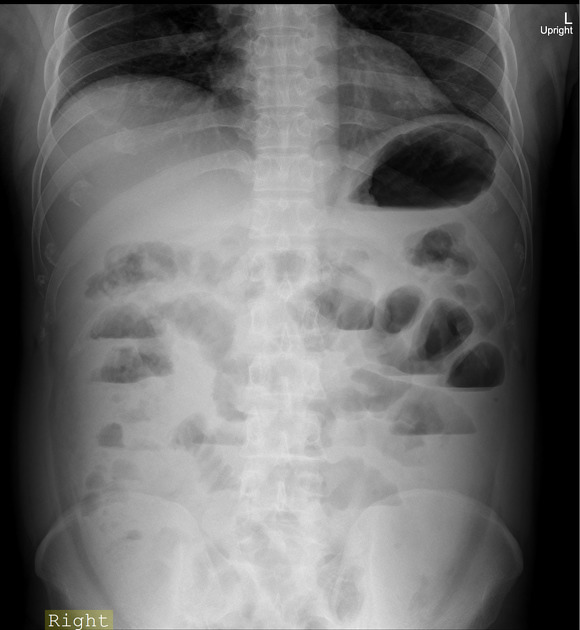 (
Image Source
)
Questions
- What are the two most probable differential diagnoses for acute mechanical bowel obstruction in this patient?
- Which initial radiological investigations should be performed to confirm the diagnosis and distinguish between these two conditions?
- List three common neonatal gastrointestinal manifestations seen in cystic fibrosis.
- What is the standard non-surgical management protocol for uncomplicated distal intestinal obstruction syndrome (DIOS) in a hemodynamically stabilized child?
Answer
- Primary Differential Diagnoses:
- Distal Intestinal Obstruction Syndrome (DIOS) (impaction of thick, viscid, inspissated mucofeculent material at the terminal ileum and cecum).
- Intussusception (ileocecal intussusception with an inspissated fecal mass acting as the pathological lead point).
- Initial Radiological Investigations:
- Abdominal ultrasonography: Evaluates for the pathognomonic "target" or "donut/pseudokidney" sign characteristic of intussusception versus an intraluminal fecal mass without telescoping.
- Water-soluble contrast enema (e.g., dilute Gastrografin / Meglumine diatrizoate): Both diagnostic (demonstrates cecal filling defect or intussusception meniscus) and potentially therapeutic (hyperosmolar agent breaks down inspissated fecal material).
- Neonatal Gastrointestinal Manifestations:
- Meconium ileus (simple or complex with volvulus/atresia)
- Meconium plug syndrome
- Meconium peritonitis (due to antenatal bowel perforation)
- Prolonged neonatal conjugated hyperbilirubinemia (cholestasis / inspissated bile syndrome)
- Non-Surgical Management Protocol for DIOS:
- Hydration: Intravenous isotonic fluid resuscitation to correct dehydration and electrolyte imbalances; complete cessation of oral intake (bowel rest) and nasogastric decompression.
- Osmotic / Fecal Lytic Agents:
- Oral or nasogastric administration of Polyethylene glycol (PEG) electrolyte solution at $1\text{ to }2\text{ g/kg/day}$ (or $20\text{ to }40\text{ mL/kg/hour}$, maximum $1\text{ to }2\text{ L/day}$) if partial obstruction and no emesis.
- In complete DIOS without bowel perforation: Gastrografin (hyperosmolar water-soluble contrast) administered orally or via therapeutic retention enema under fluoroscopic supervision with adequate intravenous pre-hydration.
RES025 - Antitubercular Drug Resistance Definitions
Scenario
A 12-year-old boy diagnosed with severe cavitary pulmonary tuberculosis has persistent sputum smear positivity at month 3 of standard first-line anti-tubercular therapy (2HRZE / 4HRE). Cartridge-Based Nucleic Acid Amplification Test (CBNAAT / GeneXpert) and line probe assay (LPA) are sent to assess for drug resistance mutations. The postgraduate trainee must categorize the susceptibility results based on updated World Health Organization (WHO) and National Tuberculosis Elimination Program (NTEP) definitions.
Questions
- Define Isoniazid-resistant TB (Hr-TB).
- Define Mono-resistant TB (MR-TB) and Poly-resistant TB (PDR-TB).
- Define Multidrug-resistant TB (MDR-TB).
- Define Extensively drug-resistant TB (XDR-TB) according to the revised 2021 WHO consensus definitions.
- Define Rifampicin-resistant TB (RR-TB) and state the clinical action required when RR-TB is identified.
Answer
- Isoniazid-Resistant Tuberculosis (Hr-TB):
- Mycobacterium tuberculosis strain resistant to Isoniazid (H), with confirmed susceptibility to Rifampicin (R).
- Mono-Resistant and Poly-Resistant Tuberculosis:
- Mono-resistant TB (MR-TB): Resistance to only one first-line anti-TB drug (e.g., isolated Isoniazid, Rifampicin, Pyrazinamide, Ethambutol, or Streptomycin resistance).
- Poly-resistant TB (PDR-TB): Resistance to more than one first-line anti-TB drug, other than both Isoniazid and Rifampicin simultaneously (e.g., resistance to Isoniazid and Ethambutol, or Pyrazinamide and Ethambutol, while retaining Rifampicin susceptibility).
- Multidrug-Resistant Tuberculosis (MDR-TB):
- Resistance to at least both Isoniazid (H) and Rifampicin (R) simultaneously, with or without resistance to other anti-TB drugs.
- Extensively Drug-Resistant Tuberculosis (XDR-TB - WHO 2021 Definition):
- Mycobacterium tuberculosis strain that meets the definition of MDR-TB or RR-TB, and is also resistant to:
- Any Fluoroquinolone (e.g., Levofloxacin or Moxifloxacin), AND
- At least one additional Group A second-line anti-TB drug (Bedaquiline or Linezolid).
- Mycobacterium tuberculosis strain that meets the definition of MDR-TB or RR-TB, and is also resistant to:
- Rifampicin-Resistant Tuberculosis (RR-TB) and Clinical Action:
- Definition: Resistance to Rifampicin detected using phenotypic or genotypic methods, with or without resistance to other anti-TB agents.
- Clinical Action: Patients with RR-TB are ineligible for standard first-line regimens and must be managed and treated with a full second-line MDR-TB treatment regimen, because the vast majority (>90%) of RR-TB strains harbor concomitant Isoniazid resistance.
More Details
graph TD
    A[Mycobacterium tuberculosis Isolated] --> B{Rifampicin Susceptibility}
    B -->|Rifampicin Susceptible| C{Isoniazid Resistance?}
    C -->|No| D[Drug-Susceptible TB]
    C -->|Yes| E[Hr-TB: Isoniazid-Resistant TB]
    B -->|Rifampicin Resistant| F[RR-TB / MDR-TB]
    F --> G{Fluoroquinolone Resistance?}
    G -->|No| H[MDR/RR-TB Standard Regimen]
    G -->|Yes| I{Group A Drug Resistance? Bedaquiline or Linezolid}
    I -->|No| J[Pre-XDR-TB]
    I -->|Yes| K[XDR-TB WHO 2021 Definition]
Note: Pre-XDR-TB is defined as MDR/RR-TB that is also resistant to any fluoroquinolone (Levofloxacin or Moxifloxacin).
RES026 - Postoperative Infant Respiratory Distress
Scenario
An 8-month-old infant is brought to the pediatric emergency department with tachypnea, grunting, and intercostal retractions. He was born at 39 weeks via vacuum-assisted vaginal delivery for shoulder dystocia, complicated by a severe left brachial plexus birth injury (flaccid left upper limb). Two months ago (at 6 months of age), he underwent left brachial plexus exploration and nerve grafting via a supraclavicular approach. On physical examination, respiratory rate is 64/min, $\text{SpO}_2$ is 88% on room air, and breath sounds are markedly diminished over the left hemithorax with paradoxical abdominal inward movement during inspiration. An urgent chest radiograph is obtained.
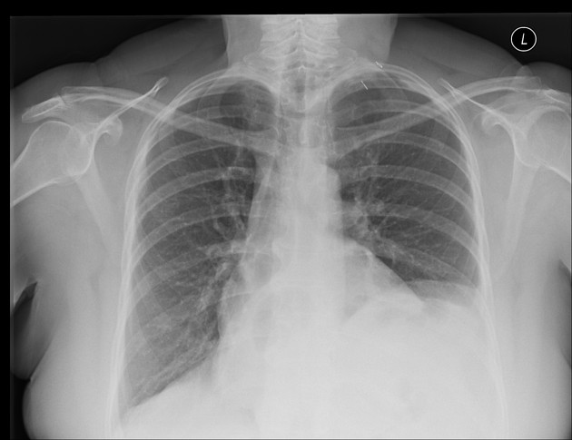 (
Image Source
)
Questions
- Enumerate three distinct radiological abnormalities expected on this patient's chest radiograph.
- What is the most likely etiology of this infant's acute respiratory decompensation?
- Name two confirmatory diagnostic modalities to evaluate dynamic diaphragmatic motion.
- Outline the definitive surgical management if this infant fails conservative management.
Answer
- Radiological Abnormalities:
- Marked elevation/eventration of the left hemidiaphragm (reaching the level of the 3rd or 4th anterior intercostal space).
- Mediastinal and tracheal shift toward the contralateral (right) side.
- Left lower lobe compressive atelectasis/collapse with surgical clips or evidence of prior clavicular osteotomy/callus formation in the supraclavicular fossa.
- Most Likely Etiology:
- Iatrogenic left phrenic nerve injury (phrenic nerve neurotmesis/traction injury) sustained during the supraclavicular neurosurgical brachial plexus reconstruction, leading to left diaphragmatic eventration/palsy.
- Confirmatory Diagnostic Modalities:
- Real-time dynamic bedside ultrasonography of the diaphragm (demonstrating paradoxical cephalad movement of the left dome during inspiration and absence of normal diaphragmatic thickening).
- Fluoroscopic sniff test (showing paradoxical upward excursion of the paralyzed left hemidiaphragm during rapid inspiration).
- Definitive Surgical Management:
- Left diaphragmatic plication (via open transthoracic, video-assisted thoracoscopic surgery [VATS], or laparoscopy) to flatten and stabilize the flaccid diaphragm, preventing paradoxical motion and restoring lung volume.
More Details
In young infants, diaphragmatic paralysis is poorly tolerated compared to older children because:
- The infant rib cage is highly compliant, offering little chest wall rigidity.
- The mediastinum is mobile, shifting markedly during paradoxical diaphragmatic excursion (pendelluft phenomenon).
- The diaphragm is the primary muscle of inspiration in early infancy.
RES027 - Pediatric Tubercular Chemotherapy Principles
Scenario
A 4-year-old child weighing 14 kg is brought to the pediatric chest clinic with low-grade fever for 3 weeks, unremitting cough, and weight loss. History reveals that the paternal grandmother passed away 2 months ago from pulmonary tuberculosis. Physical examination demonstrates severe pallor and a weight below the 3rd percentile. Gastric aspirates confirm Mycobacterium tuberculosis susceptible to all first-line drugs. You are initiating therapy per the National Tuberculosis Elimination Program (NTEP) / WHO pediatric guidelines.
Questions
- Classify the four distinct bacillary subpopulations of Mycobacterium tuberculosis in tuberculous lesions and name the primary bactericidal drug active against each.
- Formulate the standard weight-based first-line antitubercular treatment (ATT) regimen, including phases, drug names, and daily dosing (in mg/kg/day).
- Calculate the exact daily dose in milligrams for this 14 kg child for each first-line agent.
- Which micronutrient must be co-prescribed to prevent drug-induced peripheral neurotoxicity, and at what daily dose?
Answer
- Bacillary Subpopulations and Active Drugs:
- Group 1: Actively multiplying extracellular bacilli in cavity walls with neutral/alkaline pH $\to$ Isoniazid (H) (highest early bactericidal activity).
- Group 2: Slowly multiplying intracellular bacilli within macrophages in an acidic environment $\to$ Pyrazinamide (Z).
- Group 3: Slowly/intermittently multiplying semi-dormant extracellular bacilli in solid caseous necrosis ("spurters") $\to$ Rifampicin (R) (highest sterilizing capacity).
- Group 4: Completely dormant/latent bacilli within fibrotic calcified tissue $\to$ Not eradicated by active medications; suppressed by host immunity.
- Standard Pediatric First-Line ATT Regimen:
- Intensive Phase (2 months - 2 HRZE):
- Isoniazid (H): 10 mg/kg/day (range 7–15 mg/kg/day)
- Rifampicin (R): 15 mg/kg/day (range 10–20 mg/kg/day)
- Pyrazinamide (Z): 30–35 mg/kg/day (range 30–40 mg/kg/day)
- Ethambutol (E): 20 mg/kg/day (range 15–25 mg/kg/day)
- Continuation Phase (4 months - 4 HRE):
- Isoniazid (H), Rifampicin (R), and Ethambutol (E) at above daily doses.
- Intensive Phase (2 months - 2 HRZE):
- Mathematical Derivation of Daily Doses: $$\begin{aligned} \text{Child Weight} &= 14\text{ kg} \\ \text{Isoniazid (H)} &= 14\text{ kg} \times 10\text{ mg/kg/day} = \mathbf{140\text{ mg/day}} \\ \text{Rifampicin (R)} &= 14\text{ kg} \times 15\text{ mg/kg/day} = \mathbf{210\text{ mg/day}} \\ \text{Pyrazinamide (Z)} &= 14\text{ kg} \times 35\text{ mg/kg/day} = \mathbf{490\text{ mg/day}} \\ \text{Ethambutol (E)} &= 14\text{ kg} \times 20\text{ mg/kg/day} = \mathbf{280\text{ mg/day}} \end{aligned} $$
- Micronutrient Supplementation:
- Pyridoxine (Vitamin B6): 10 mg/day orally (administered to infants, malnourished children, HIV-infected children, and exclusively breastfed infants to counter INH-mediated pyridoxine antagonism).
RES028 - Drug-Resistant Tuberculosis Regimen Formulation
Scenario
A 6-year-old child weighing 18 kg is diagnosed with microbiologically confirmed pulmonary tuberculosis. Rapid molecular testing via cartridge-based nucleic acid amplification test (CBNAAT) and subsequent first-line line probe assay (FL-LPA) reveals Mycobacterium tuberculosis with a katG mutation conferring high-level resistance to Isoniazid, while Rifampicin resistance is not detected (Rifampicin-sensitive). You are tasked with initiating the national evidence-based regimen for Isoniazid mono-resistant tuberculosis (Hr-TB).
Questions
- Specify the recommended drug regimen and its minimum standard duration for Isoniazid mono-resistant tuberculosis according to NTEP / WHO guidelines.
- Calculate the specific daily dose (in mg) for each drug in this regimen for this 18 kg child.
- Under what clinical or radiological circumstance is the duration of this regimen prolonged, and to what total duration?
- What mandatory baseline sensory assessment must be documented prior to initiating the fluoroquinolone and ethambutol components?
Answer
- Recommended Regimen & Duration:
- Regimen: 6 (Lfx - R - Z - E)
- Daily administration of Levofloxacin, Rifampicin, Pyrazinamide, and Ethambutol for a duration of 6 months.
- Daily Drug Dosing: $$\begin{aligned} \text{Child Weight} &= 18\text{ kg (age } \ge 5\text{ years)} \\ \text{Levofloxacin (10--15 mg/kg/day)} &= 18\text{ kg} \times 10\text{ to } 15\text{ mg/kg} = \mathbf{180\text{ to } 270\text{ mg/day}} \text{ (typically 250 mg once daily)} \\ \text{Rifampicin (15 mg/kg/day)} &= 18\text{ kg} \times 15\text{ mg/kg} = \mathbf{270\text{ mg/day}} \\ \text{Pyrazinamide (30--35 mg/kg/day)} &= 18\text{ kg} \times 35\text{ mg/kg} = \mathbf{630\text{ mg/day}} \\ \text{Ethambutol (20 mg/kg/day)} &= 18\text{ kg} \times 20\text{ mg/kg} = \mathbf{360\text{ mg/day}} \end{aligned} $$
- Indication for Prolongation:
- In extensive cavitary pulmonary disease, bilateral parenchymal destruction, or slow smear/culture conversion at month 2–3, the regimen duration is extended from 6 months to 9 months.
- Mandatory Baseline Assessments:
- Visual assessment: Visual acuity and color vision testing (Ishihara plates) to screen for ethambutol-induced retrobulbar neuritis.
- Cardiac electrophysiology: Baseline 12-lead electrocardiogram (ECG) to determine corrected QT interval ($\text{QTc}$) prior to initiating Levofloxacin.
RES029 - Congenital Intranasal Mass Evaluation
Scenario
A 6-month-old infant is evaluated in the pediatric outpatient clinic for progressive unilateral nasal obstruction, intermittent stertor, and difficulty during bottle feeding. Anterior rhinoscopy demonstrates a smooth, pinkish, non-ulcerated mass within the left nasal cavity. The pediatric resident considers a congenital midline mass versus an inflammatory lesion.
Questions
- Match each lesion (A through D) with its characteristic clinical or structural findings (1 through 5; options may be used more than once or not at all):
- Lesions:
- A. Intranasal polyp
- B. Nasofrontal/nasoethmoidal encephalocele
- C. Nasal glioma (heterotopic neuroglial tissue)
- D. Nasal dermoid sinus cyst
- Characteristics:
- Strictly intranasal; never possesses an extranasal external component
- Expands on crying/straining, compressible, and demonstrates transillumination
- Obligatory cranial bony defect with direct intracranial subarachnoid communication
- Non-pulsatile, firm, no expansion on jugular compression, isolated glial rests
- Pit or sinus opening on midline nasal dorsum with protruding hair
- Lesions:
- What is the classic bedside maneuver used to evaluate intracranial communication of an intranasal mass?
- What is the gold-standard imaging strategy required before any tissue biopsy or surgical intervention?
- What serious clinical complication can occur if a congenital encephalocele is biopsied without pre-procedural neuroimaging?
Answer
- Matching Lesions to Characteristics:
- A. Intranasal polyp $\to$ 1 (Strictly intranasal; never possesses an extranasal external component).
- B. Encephalocele $\to$ 2, 3 (Expands on crying, transilluminates; obligatory cranial defect with direct CSF communication).
- C. Nasal glioma $\to$ 4 (Non-pulsatile, firm, no expansion on jugular compression, isolated glial rests without persistent patent CSF tract in ~85%).
- D. Nasal dermoid sinus cyst $\to$ 5 (Midline nasal dorsal pit/ostium, frequently with protruding hair or sebaceous discharge).
- Furstenberg Test:
- Compression of both internal jugular veins causes engorgement and expansion/pulsation of the intranasal mass in an encephalocele due to increased intracranial venous pressure; a glioma or polyp shows no change.
- Gold-Standard Imaging:
- High-resolution thin-cut (0.5–1 mm) computed tomography (HRCT) of the skull base and facial bones (to delineate the bony defect at the cribriform plate/foramen cecum) paired with high-resolution magnetic resonance imaging (MRI) of the brain and orbits (to demonstrate neural tissue herniation and CSF continuity).
- Complication of Inadvertent Biopsy:
- Direct cerebrospinal fluid (CSF) leak, ascending bacterial meningitis, ventriculitis, and brain abscess.
RES030 - Mechanical Ventilation Graphic Interpretation
Scenario
A 3-day-old preterm infant born at 28 weeks of gestation is receiving synchronized intermittent mandatory ventilation with volume guarantee (SIMV-VG) in the neonatal intensive care unit for persistent respiratory failure. The following pressure-volume (P-V) loop is displayed on the ventilator monitor:
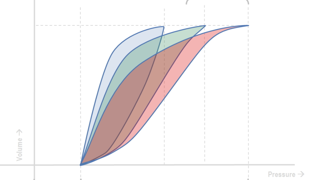 (
Image Source
)
Questions
- Describe the three key graphic abnormalities observed in this pressure-volume (P-V) loop (red loop).
- What underlying mechanical lung abnormality does this graphic loop signify?
- Calculate the static respiratory system compliance ($\text{C}_{\text{stat}}$) if the delivered tidal volume ($\text{V}_{\text{t}}$) is 12 mL, plateau pressure ($\text{P}_{\text{plat}}$) is 28 $\text{cmH}_2\text{O}$, and positive end-expiratory pressure ($\text{PEEP}$) is 6 $\text{cmH}_2\text{O}$ in this 1.2 kg neonate. State whether this calculated value is normal, increased, or decreased.
- Enumerate three underlying pulmonary pathologies in a neonate that produce this specific P-V loop configuration.
Answer
- P-V Loop Graphic Abnormalities:
- Inspiratory limb is flattened with a sluggish upward slope, shifted downward and to the right toward the pressure axis.
- Narrow loop area reflecting small change in volume per unit change in pressure ($\Delta\text{V}/\Delta\text{P}$).
- Prominent lower inflection point (LIP) representing delayed alveolar opening/recruitment pressure.
- Underlying Mechanical Abnormality:
- Marked reduction in dynamic and static respiratory compliance (stiff, non-compliant lungs).
- Compliance Calculation:
$$\begin{aligned}
\text{C}_{\text{stat}} &= \frac{\text{V}_{\text{t}}}{\text{P}_{\text{plat}} - \text{PEEP}} \\
&= \frac{12\text{ mL}}{28\text{ cmH}_2\text{O} - 6\text{ cmH}_2\text{O}} \\
&= \frac{12}{22} = \mathbf{0.55\text{ mL/cmH}_2\text{O}} \\
\text{Normalized for weight} &= \frac{0.55\text{ mL/cmH}_2\text{O}}{1.2\text{ kg}} = \mathbf{0.46\text{ mL/cmH}_2\text{O/kg}}
\end{aligned}
$$
- Interpretation: Markedly decreased (Normal neonatal respiratory compliance is $1.0\text{ to }2.0\text{ mL/cmH}_2\text{O/kg}$).
- Underlying Causes:
- Respiratory Distress Syndrome (Surfactant deficiency / Hyaline Membrane Disease).
- Pulmonary hemorrhage.
- Tension or significant pneumothorax.
- Severe pulmonary edema or Congenital Diaphragmatic Hernia with pulmonary hypoplasia.
RES031 - Pediatric Mechanical Ventilation Parameter Adjustments
Scenario
A 5-year-old child weighing 18 kg with severe community-acquired pneumonia is intubated and mechanically ventilated in the Pediatric Intensive Care Unit (PICU). The ventilator is functioning in a time-cycled, pressure-limited mode with the following initial baseline settings:
- Peak Inspiratory Pressure (PIP): $18\text{ cmH}_2\text{O}$
- Positive End-Expiratory Pressure (PEEP): $5\text{ cmH}_2\text{O}$
- Set Respiratory Rate (RR): $20\text{ breaths/min}$
- Inspiratory-to-Expiratory (I:E) Ratio: $1:2$
- Fraction of Inspired Oxygen ($\text{FiO}_2$): $0.40$
An arterial blood gas (ABG) drawn 30 minutes after stabilization reveals:
- $\text{pH}: 7.24$
- $\text{PaCO}_2: 70\text{ mmHg}$
- $\text{PaO}_2: 94\text{ mmHg}$
- $\text{HCO}_3^-: 29\text{ mEq/L}$
- $\text{SaO}_2: 97\%$
Questions
- Identify the primary ventilatory problem and state the two most effective adjustments in ventilator settings to achieve normocapnia.
- Calculate the total respiratory cycle time ($T_{\text{tot}}$), inspiratory time ($T_i$), and expiratory time ($T_e$) for this child based on current settings.
- Calculate the target minute ventilation or respiratory rate required to bring the $\text{PaCO}_2$ down to $40\text{ mmHg}$ (assuming constant $\text{CO}_2$ production and dead space).
- Define Continuous Positive Airway Pressure (CPAP) and explain how it differs from Bi-level Positive Airway Pressure (BiPAP) in non-invasive respiratory support.
Answer
- Primary Ventilatory Defect and Setting Adjustments:
- Primary Problem: Severe alveolar hypoventilation leading to acute-on-chronic respiratory acidosis with hypercapnia, in the presence of adequate oxygenation ($\text{PaO}_2 > 80\text{ mmHg}$, $\text{SaO}_2 > 95\%$).
- Adjustments to Normalize $\text{PaCO}_2$:
- Increase Ventilator Respiratory Rate (RR): Directly increases minute ventilation ($V_E = \text{RR} \times V_T$) without increasing peak airway pressures or barotrauma risk.
- Increase Peak Inspiratory Pressure (PIP): Increases driving pressure ($\Delta P = \text{PIP} - \text{PEEP}$), thereby expanding tidal volume ($V_T$) and augmenting alveolar ventilation.
- Ventilatory Timing Calculations: $$\begin{aligned} T_{\text{tot}} &= \frac{60\text{ seconds}}{\text{Set RR}} = \frac{60}{20} = \mathbf{3.0\text{ seconds}} \\ \text{Sum of parts in I:E ratio } (1:2) &= 1 + 2 = 3 \\ T_i &= \frac{1}{3} \times T_{\text{tot}} = \frac{1}{3} \times 3.0 = \mathbf{1.0\text{ second}} \\ T_e &= \frac{2}{3} \times T_{\text{tot}} = \frac{2}{3} \times 3.0 = \mathbf{2.0\text{ seconds}} \end{aligned} $$
- Mathematical Derivation for New Respiratory Rate: $$\begin{aligned} \text{Target RR} &= \frac{\text{Current RR} \times \text{Current }\text{PaCO}_2}{\text{Target }\text{PaCO}_2} \\ &= \frac{20 \times 70}{40} \\ &= \frac{1400}{40} = \mathbf{35\text{ breaths/min}} \end{aligned} $$ (Alternatively, increasing tidal volume proportionally by increasing PIP from $18\text{ cmH}_2\text{O}$ while maintaining rate).
- CPAP versus BiPAP:
- Continuous Positive Airway Pressure (CPAP): Delivers a constant predetermined positive airway pressure throughout both inspiration and expiration during spontaneous breathing. It recruits atelectatic alveoli, increases Functional Residual Capacity (FRC), and improves oxygenation without providing active inspiratory pressure support.
- Bi-level Positive Airway Pressure (BiPAP): Delivers two distinct pressure levels—an Inspiratory Positive Airway Pressure (IPAP) to assist tidal ventilation and clear $\text{CO}_2$, and an Expiratory Positive Airway Pressure (EPAP) to maintain FRC and oxygenation.
RES032 - Childhood Orthopnea And Thoracic Opacity
Scenario
An 8-year-old boy presents to the emergency room with progressive dyspnea, dry cough, facial puffiness, and inability to lie flat (severe orthopnea) over the past 2 weeks. On examination, he is tachypneic (RR 44/min) with prominent suprasternal and subcostal retractions. He has marked pallor, generalized lymphadenopathy (bilateral cervical and axillary nodes measuring $1.5 \times 1.5\text{ cm}$, firm, non-tender, discrete), hepatomegaly of 4 cm below the right costal margin, and splenomegaly of 5 cm below the left costal margin.
An urgent erect portable chest radiograph is performed as part of the initial evaluation.
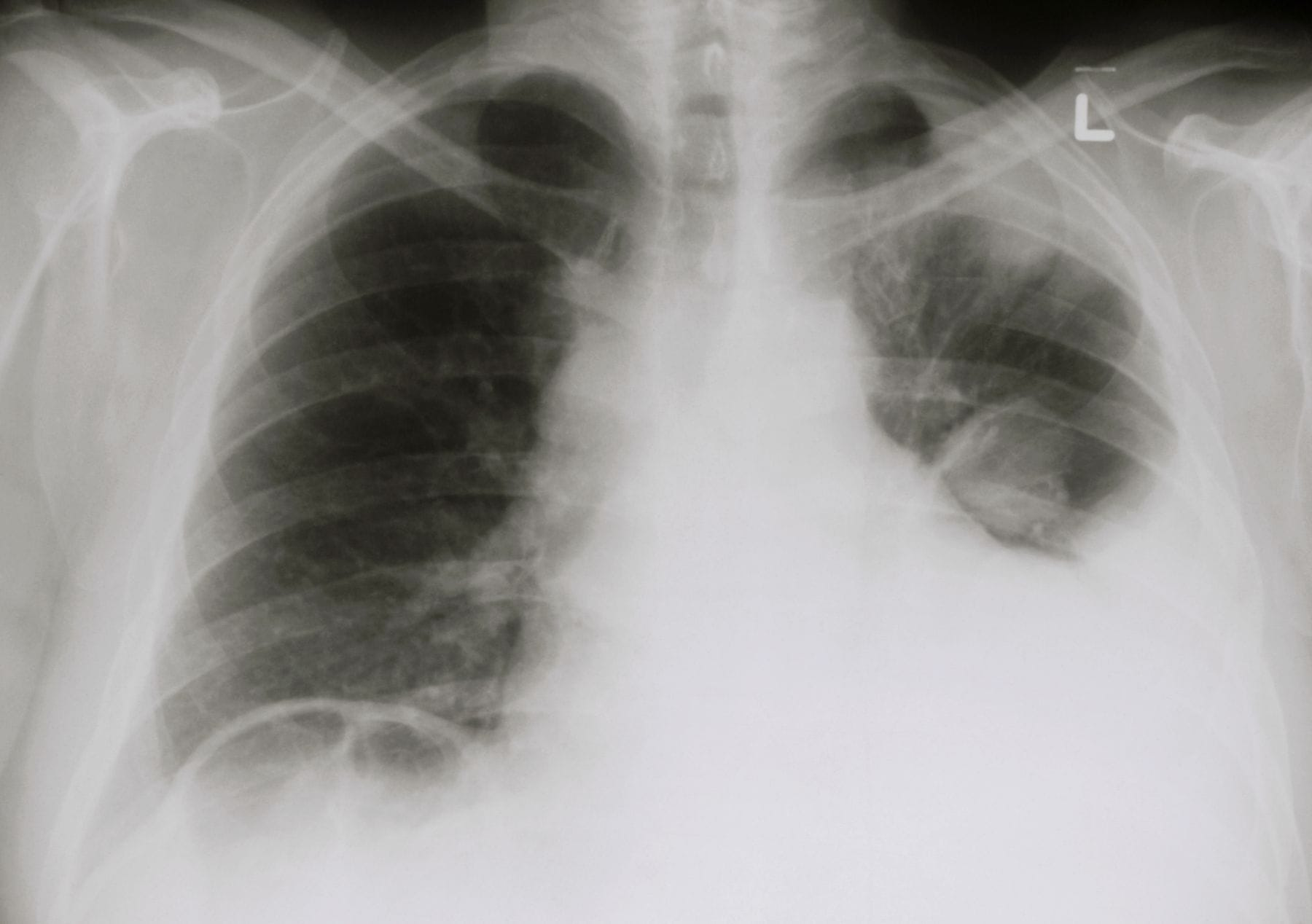 (
Image Source
)
Questions
- Describe four salient abnormal radiological findings on the provided chest radiograph.
- What is the most probable clinical and radiological diagnosis?
- Enumerate the immediate life-threatening physiological complication (syndrome) this child is at imminent risk of developing.
- List four critical precautions/contraindications during the emergency stabilization and induction of anesthesia in this patient.
- Outline three diagnostic investigations required to confirm the primary etiology without compromising patient safety.
Answer
- Radiological Findings:
- Homogeneous dense radio-opacity occupying the left hemithorax with obliteration of the left costophrenic and cardiophrenic angles (massive left-sided pleural effusion).
- Significant shift of the mediastinum, trachea, and cardiac silhouette towards the contralateral (right) side.
- Widening of the superior anterior mediastinum visible above the effusion shadow.
- Presence of an intercostal chest drainage tube in situ on the left hemithorax.
- Most Probable Diagnosis:
- Anterior mediastinal mass with massive left-sided exudative/malignant pleural effusion, most likely secondary to T-cell Acute Lymphoblastic Leukemia / Lymphoblastic Lymphoma (T-ALL / T-LBL).
- Life-Threatening Complication:
- Superior Mediastinal Syndrome (SMS) / Superior Vena Cava (SVC) Syndrome accompanied by critical tracheobronchial compression and acute cardiorespiratory collapse.
- Critical Precautions during Stabilization:
- Avoid Supine Positioning: Keep the child in an upright, orthopneic, or prone/lateral position of comfort to prevent complete airway collapse.
- Avoid General Anesthesia and Neuromuscular Blockade: Paralysis eliminates spontaneous negative-pressure ventilation and dynamic airway tone, causing fatal, irreversible tracheobronchial compression.
- Avoid Sedation / Anxiolytics: Sedation suppresses respiratory drive and compensatory accessory muscle use.
- Controlled Pleural Decompression: Avoid rapid, high-volume pleural evacuation to prevent re-expansion pulmonary edema and sudden cardiovascular shift.
- Diagnostic Investigations:
- Peripheral Blood Smear & Flow Cytometry: To detect circulating lymphoblasts without invasive tissue biopsy.
- Diagnostic Pleural Fluid Aspiration (Local Anesthesia): Cytospin, cell count, biochemistry, and immunophenotyping/flow cytometry of pleural fluid.
- Peripheral Lymph Node Biopsy: Excisional or core biopsy of a superficial cervical/axillary node under local anesthesia.
- Bone Marrow Aspirate/Biopsy: Under local anesthesia in a non-supine position if peripheral blasts are absent.
RES033 - Persistent Fever And Mediastinal Lymphadenopathy
Scenario
A 10-year-old girl is evaluated for continuous low-grade fever with evening worsening, poor appetite, unquantified weight loss, and an intermittent dry hacking cough lasting for 6 weeks. There is no history of hemoptysis. A tuberculin skin test (Mantoux) reveals $18\text{ mm}$ induration at 72 hours. Contrast-Enhanced Computed Tomography (CECT) of the chest is performed to investigate persistent mediastinal widening on plain chest radiography.
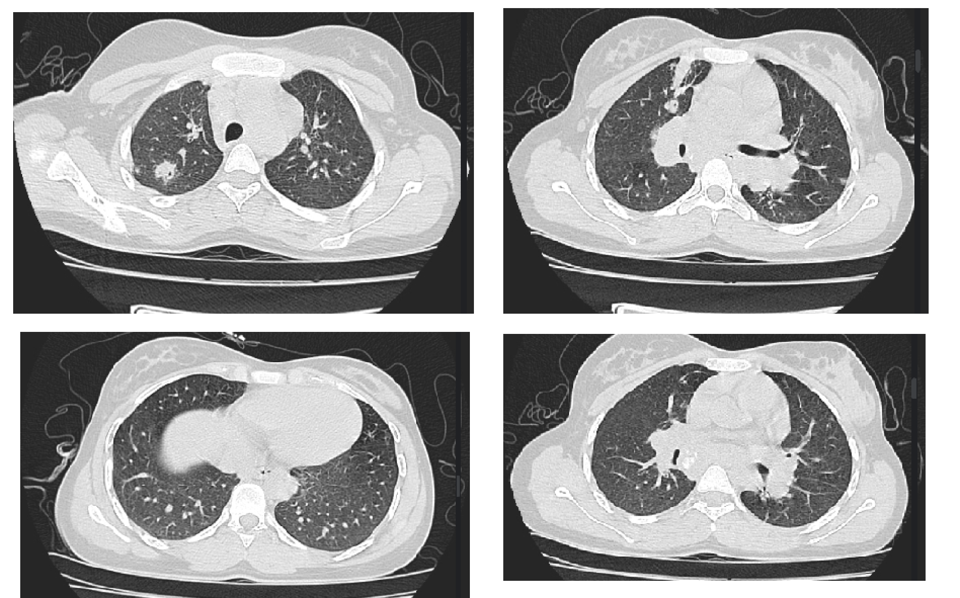 (
Image Source
)
Questions
- Describe the characteristic imaging findings on the provided CECT chest.
- What is the most probable diagnosis based on the imaging features?
- Mention three close differential diagnoses for these imaging findings in a pediatric patient.
- What is the preferred minimally invasive modality to obtain tissue/microbiological confirmation if sputum/gastric aspirates are negative?
- State the standard first-line treatment regimen under the National Tuberculosis Elimination Program (NTEP) including drugs, phases, and duration.
Answer
- Characteristic CECT Chest Findings:
- Multiple enlarged mediastinal, paratracheal, pretracheal, subcarinal, and hilar lymph nodes.
- Nodes demonstrate characteristic central low-attenuation (hypodense) areas representing caseous necrosis.
- Prominent peripheral rim-like contrast enhancement (ring-enhancing nodal masses).
- Compression or luminal narrowing of adjacent major tracheobronchial structures.
- Most Probable Diagnosis:
- Mediastinal Tuberculous Lymphadenitis (Thoracic Tuberculosis).
- Differential Diagnoses:
- Lymphoma (Hodgkin or Non-Hodgkin Lymphoma—typically homogeneous enhancement without central necrosis prior to therapy).
- Fungal Lymphadenitis (Histoplasmosis, Coccidioidomycosis).
- Sarcoidosis (typically non-necrotic, symmetric bilateral hilar and right paratracheal lymphadenopathy).
- Metastatic Lymphadenopathy (e.g., metastatic embryonal rhabdomyosarcoma, neuroblastoma).
- Preferred Modality for Microbiological Confirmation:
- Endobronchial Ultrasound-guided Transbronchial Needle Aspiration (EBUS-TBNA) or Endoscopic Ultrasound (EUS-FNA) for smear microscopy, Cartridge Based Nucleic Acid Amplification Test (CBNAAT / GeneXpert MTB/RIF), and mycobacterial culture.
- Standard NTEP First-Line Treatment Regimen:
- Intensive Phase (2 months - 2HRZE):
- Isoniazid (H): $10\text{ mg/kg/day}$ (range 7–15 mg/kg)
- Rifampicin (R): $15\text{ mg/kg/day}$ (range 10–20 mg/kg)
- Pyrazinamide (Z): $35\text{ mg/kg/day}$ (range 30–40 mg/kg)
- Ethambutol (E): $20\text{ mg/kg/day}$ (range 15–25 mg/kg)
- Continuation Phase (4 months - 4HRE):
- Isoniazid (H), Rifampicin (R), and Ethambutol (E) daily for 16 weeks.
- Total duration: 6 months of daily, weight-band based, fixed-dose combination (FDC) therapy.
- Intensive Phase (2 months - 2HRZE):
RES034 - Pediatric Shock Acid Base Derangement
Scenario
A 4-year-old boy weighing 15 kg is brought to the pediatric emergency department with acute lethargy, persistent vomiting, and poor oral intake for 2 days. On examination, he is obtunded with deep, rapid respirations (Kussmaul breathing, RR 42/min). His peripheral extremities are cold and mottled, radial pulses are non-palpable, femoral pulses are weak, capillary refill time is 5 seconds, heart rate is 172 beats/min, and blood pressure is $72/44\text{ mmHg}$.
An emergency arterial blood gas (ABG) and serum biochemistry drawn simultaneously reveal:
- $\text{pH}: 7.15$
- $\text{PaCO}_2: 25\text{ mmHg}$
- $\text{PaO}_2: 90\text{ mmHg}$ (on room air)
- $\text{HCO}_3^-: 11\text{ mEq/L}$
- $\text{Base Deficit}: -15\text{ mEq/L}$
- $\text{Serum Sodium } (\text{Na}^+): 134\text{ mEq/L}$
- $\text{Serum Potassium } (\text{K}^+): 4.8\text{ mEq/L}$
- $\text{Serum Chloride } (\text{Cl}^-): 98\text{ mEq/L}$
- $\text{Blood Glucose}: 86\text{ mg/dL}$
- $\text{Serum Lactate}: 7.2\text{ mmol/L}$
Questions
- Provide the complete structured interpretation of the arterial blood gas.
- Calculate the serum anion gap and evaluate respiratory compensation using Winter's formula.
- What is the underlying pathophysiological cause for this acid-base disturbance?
- Specify the immediate crystalloid fluid resuscitation protocol (fluid choice, volume in mL, and infusion rate).
- State the evidence-based recommendation regarding intravenous sodium bicarbonate administration in this patient.
Answer
- Arterial Blood Gas Interpretation:
- Primary Disorder: Fully compensated severe metabolic acidosis.
- Secondary Physiological Response: Secondary compensatory respiratory alkalosis (hyperventilation/Kussmaul breathing).
- Oxygenation: Adequate arterial oxygenation on room air ($\text{PaO}_2 = 90\text{ mmHg}$).
- Mathematical Calculations:
$$\begin{aligned}
\text{Serum Anion Gap} &= [\text{Na}^+] - ([\text{Cl}^-] + [\text{HCO}_3^-]) \\
&= 134 - (98 + 11) \\
&= 134 - 109 = \mathbf{25\text{ mEq/L}} \quad (\text{Normal: } 8\text{--}12\text{ mEq/L}) \\
&\implies \text{High Anion Gap Metabolic Acidosis (HAGMA)}
\end{aligned}
$$
$$\begin{aligned}
\text{Expected }\text{PaCO}_2 \text{ (Winter's formula)} &= (1.5 \times [\text{HCO}_3^-]) + 8 \pm 2 \\
&= (1.5 \times 11) + 8 \pm 2 \\
&= 16.5 + 8 \pm 2 = \mathbf{24.5 \pm 2\text{ mmHg}} \quad (22.5\text{--}26.5\text{ mmHg})
\end{aligned}
$$
- Since measured $\text{PaCO}_2$ ($25\text{ mmHg}$) falls precisely within the expected range ($22.5\text{--}26.5\text{ mmHg}$), there is appropriate, simple respiratory compensation without a co-existing secondary respiratory disorder.
- Underlying Pathophysiology:
- Hypovolemic / Septic Shock with Tissue Hypoperfusion: Inadequate systemic oxygen delivery ($DO_2$) shifts cellular respiration to anaerobic glycolysis, generating excess lactic acid (L-lactate $7.2\text{ mmol/L}$), unmeasured organic anions, and severe metabolic acidosis.
- Immediate Fluid Resuscitation:
- Fluid Choice: Balanced isotonic crystalloid (e.g., Plasmalyte or Ringer's Lactate) or $0.9\%$ Normal Saline.
- Dose: $20\text{ mL/kg}$ bolus.
- Volume for 15 kg Child}: $20 \times 15 = \mathbf{300\text{ mL}}$.
- Infusion Rate: Administer via rapid push or pressure bag over 10 to 20 minutes, followed by reassessment of perfusion markers (pulses, CRT, mental status).
- Role of Sodium Bicarbonate:
- Contraindicated as routine initial therapy.
- Rationale: Bicarbonate combines with hydrogen ions to produce $\text{CO}_2$ ($\text{H}^+ + \text{HCO}_3^- \rightleftharpoons \text{H}_2\text{CO}_3 \rightleftharpoons \text{H}_2\text{O} + \text{CO}_2$). $\text{CO}_2$ freely diffuses across cell membranes and the blood-brain barrier faster than bicarbonate, exacerbating paradoxical intracellular and cerebral acidosis, shifting the oxyhemoglobin dissociation curve to the left (reducing tissue oxygen release), and inducing hypocalcemia and hypernatremia.
- Definitive treatment: Restoration of intravascular volume and tissue perfusion via crystalloid boluses to clear lactate.
RES035 - Diffuse Micronodular Pulmonary Infiltrates Assessment
Scenario
A 6-year-old boy presents with progressive low-grade fever, anorexia, night sweats, and worsening dyspnea for 5 weeks. He has not received the BCG vaccine. On physical examination, he appears chronically ill, wasted, and tachypneic. Auscultation of the chest reveals bilateral diffuse fine end-inspiratory crackles. Fundoscopic examination reveals bilateral pale-yellow choroidal tubercles. High-Resolution Computed Tomography (HRCT) of the chest is shown below.
 (
Image Source
)
Questions
- Describe the key radiological findings on the provided HRCT chest scan.
- Provide five differential diagnoses (infectious and non-infectious) that present with this radiological pattern.
- List three definitive microbiological or molecular tests to confirm the diagnosis.
- Name two critical extra-pulmonary screening evaluations that must be performed immediately.
- Detail the complete pharmacological management plan including anti-tubercular drug regimen, duration, and adjuvant therapy.
Answer
- Key HRCT Chest Findings:
- Diffuse, widespread, bilateral micronodules ($1\text{ to }3\text{ mm}$ in diameter).
- Random / Miliary distribution: Nodules show no predilection for bronchovascular bundles, centrilobular regions, or subpleural surfaces, uniformly involving all lung lobes from apex to base.
- Differential Diagnoses for Miliary / Micronodular Pattern:
- Miliary Tuberculosis (most common in endemic regions).
- Disseminated Fungal Infections (Histoplasmosis, Blastomycosis, Coccidioidomycosis).
- Langerhans Cell Histiocytosis (LCH) (nodular phase).
- Metastatic Malignancy (e.g., Papillary thyroid carcinoma, Wilms tumor, Osteosarcoma).
- Hypersensitivity Pneumonitis or Sarcoidosis (typically perilymphatic rather than random).
- Disseminated Viral Pneumonitis (e.g., Varicella, Cytomegalovirus in immunocompromised hosts).
- Definitive Diagnostic Tests:
- Gastric Aspirates (3 consecutive mornings) or Induced Sputum / BAL Fluid:
- GeneXpert MTB/RIF Ultra (CBNAAT) for rapid detection of Mycobacterium tuberculosis DNA and rifampicin resistance.
- Acid-Fast Bacilli (AFB) smear microscopy (Ziehl-Neelsen or Auramine-O fluorescence).
- Liquid mycobacterial culture (MGIT - Mycobacteria Growth Indicator Tube).
- Gastric Aspirates (3 consecutive mornings) or Induced Sputum / BAL Fluid:
- Critical Extra-pulmonary Screenings:
- Contrast-Enhanced Neuroimaging (CE-MRI / CECT Brain) and Lumbar Puncture: To screen for asymptomatic Tuberculous Meningitis (TBM) or tuberculomas (present in up to $30\text{--}40\%$ of miliary TB).
- Abdominal Ultrasonography: To identify hepatosplenic granulomas, retroperitoneal lymphadenopathy, or ascites.
- Pharmacological Management:
- Anti-Tubercular Therapy (ATT):
- Intensive Phase: 4 drugs (HRZE - Isoniazid, Rifampicin, Pyrazinamide, Ethambutol) daily for 2 months.
- Continuation Phase: 3 drugs (HRE - Isoniazid, Rifampicin, Ethambutol) daily for 7 to 10 months (Total duration: 9 to 12 months for disseminated/miliary disease).
- Adjuvant Corticosteroid Therapy:
- Prednisolone: $2\text{ mg/kg/day}$ (maximum $60\text{ mg/day}$) orally for 4 to 6 weeks, followed by a gradual taper over 2 to 4 weeks. Indicated to reduce alveolar-capillary block, treat hypoxemia, and prevent severe immune-mediated systemic inflammation.
- Anti-Tubercular Therapy (ATT):
More Details
- Random (Miliary) Pattern: Nodules are distributed uniformly without anatomical preference relative to the secondary pulmonary lobule (seen in hematogenous dissemination: TB, fungal, hematogenous metastases).
- Perilymphatic Pattern: Nodules follow lymphatic channels along pleural surfaces, interlobular septa, and peribronchovascular bundles (seen in sarcoidosis, silicosis, lymphangitic carcinomatosis).
- Centrilobular Pattern: Nodules spare the pleural surfaces and interlobular septa, staying $2\text{--}3\text{ mm}$ away from the pleural margin (seen in endobronchial spread of TB, hypersensitivity pneumonitis, bronchiolitis).
RES036 - Diffuse Micronodular Pulmonary Infiltrates
Scenario
A 14-month-old unimmunized girl is brought to the pediatric emergency department with a 4-week history of low-grade intermittent fever, poor feeding, progressive weight loss, and worsening non-productive cough. On examination, she is irritable and tachypneic. Her vitals reveal a heart rate of 142/min, respiratory rate of 56/min with mild intercostal retractions, temperature of 38.3°C (101°F), and oxygen saturation of 91% on room air. Her weight-for-age is below the 3rd percentile. Systemic examination reveals generalized nontender lymphadenopathy and firm hepatosplenomegaly (liver palpable 3.5 cm and spleen 2.5 cm below costal margins). Fine bilateral end-inspiratory crepitations are audible across all lung fields. A digital chest radiograph is obtained.
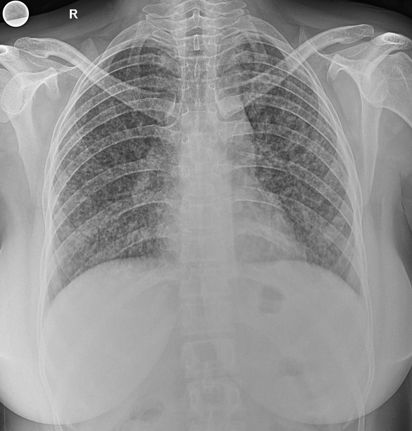 (
Image Source
)
Questions
- Describe the cardinal radiological abnormalities shown on this chest radiograph and formulate the definitive diagnosis.
- List four distinct non-tuberculous differential diagnoses that can present with an identical radiological appearance in early childhood.
- Detail the recommended National Tuberculosis Elimination Program (NTEP) / WHO weight-based anti-tubercular therapy (ATT) regimen for this patient, specifying drugs, phases, and total duration.
- Enumerate two essential non-pulmonary clinical evaluations required immediately to evaluate dissemination, and name the adjunctive pharmacological agent indicated if neurological involvement is confirmed.
Answer
- Radiological Description & Diagnosis:
- Findings: Diffuse, bilateral, uniform, discrete micronodular opacities ("millet-seed" appearance, 1–2 mm in diameter) distributed symmetrically throughout both lung fields from apex to base, with loss of sharp margin definition.
- Primary Diagnosis: Miliary Tuberculosis (Disseminated Tuberculosis).
- Differential Diagnoses:
- Disseminated fungal infection (e.g., Histoplasmosis, Coccidioidomycosis).
- Langerhans Cell Histiocytosis (Pulmonary LCH).
- Idiopathic Pulmonary Hemosiderosis.
- Sarcoidosis or Extrinsic Allergic Alveolitis (Hypersensitivity Pneumonitis).
- Metastatic malignancy (e.g., metastatic Neuroblastoma, Wilms tumor, or hepatoblastoma).
- Treatment Regimen (NTEP / WHO Pediatric Protocol):
- Intensive Phase (IP - 2 months): Four drugs daily using pediatric Fixed-Dose Combinations (FDCs):
- Isoniazid (H): $10\text{ mg/kg/day}$ (range $7\text{–}15\text{ mg/kg/day}$)
- Rifampicin (R): $15\text{ mg/kg/day}$ (range $10\text{–}20\text{ mg/kg/day}$)
- Pyrazinamide (Z): $35\text{ mg/kg/day}$ (range $30\text{–}40\text{ mg/kg/day}$)
- Ethambutol (E): $20\text{ mg/kg/day}$ (range $15\text{–}25\text{ mg/kg/day}$)
- Continuation Phase (CP - 4 to 10 months): Three drugs daily (HRE) for 4 months (total 6 months). If concurrent TB meningitis or spinal/osteoarticular disease is identified, extend continuation phase to 10 months (total 12 months duration).
- Intensive Phase (IP - 2 months): Four drugs daily using pediatric Fixed-Dose Combinations (FDCs):
- Screening & Adjunctive Therapy:
- Screening Evaluations:
- Dilated fundoscopic examination to detect choroid tubercles.
- Lumbar puncture for CSF analysis and neuroimaging (contrast-enhanced cranial MRI/CT) to rule out subclinical Tuberculous Meningitis (TBM).
- Adjunctive Pharmacotherapy: Systemic corticosteroids (Oral Prednisolone $2\text{ mg/kg/day}$ or IV Dexamethasone $0.4\text{ mg/kg/day}$, tapered gradually over 4 to 6 weeks).
- Screening Evaluations:
RES037 - Evaluation of Refractory Tuberculosis
Scenario
A 4-year-old boy with microbiologically confirmed pulmonary tuberculosis fails to show clinical and radiological improvement despite 8 weeks of adherent, directly observed first-line anti-tubercular therapy (HRZE). Sputum induction yields an acid-fast bacillus (AFB) smear-positive specimen. You plan to perform rapid molecular testing to detect comprehensive first- and second-line drug resistance mutations to configure an appropriate second-line therapeutic regimen.
Questions
- Name the rapid molecular diagnostic platform endorsed by the WHO and NTEP for detecting resistance to first-line and second-line anti-tubercular agents, and state the prerequisite specimen criteria.
- State the turnaround time for this assay and list the specific genetic loci evaluated to determine resistance to Isoniazid, Rifampicin, and Fluoroquinolones.
- Provide the updated WHO definitions for Multidrug-Resistant TB (MDR-TB), Pre-Extensively Drug-Resistant TB (Pre-XDR-TB), and Extensively Drug-Resistant TB (XDR-TB).
- Outline the composition of an all-oral longer regimen for pediatric MDR/RR-TB, classifying agents according to WHO Groups A, B, and C.
Answer
- Diagnostic Test & Specimen Requirement:
- Test: Line Probe Assay (LPA) (e.g., GenoType MTBDRplus for first-line agents and GenoType MTBDRsl for second-line agents).
- Prerequisite: Direct AFB smear-positive clinical specimen or an active culture isolate of Mycobacterium tuberculosis.
- Turnaround Time & Genetic Targets:
- Turnaround Time: $48\text{ to }72\text{ hours}$.
- Genetic Targets:
- Rifampicin resistance: Mutations in the 81-base-pair Rifampicin Resistance Determining Region (RRDR) of the $rpoB$ gene.
- Isoniazid resistance: High-level resistance via $katG$ gene mutations (codon 315); low-level resistance via promoter region mutations of the $inhA$ gene.
- Fluoroquinolone resistance: Mutations in the quinolone resistance-determining regions (QRDR) of the $gyrA$ and $gyrB$ genes.
- Case Definitions (WHO 2021 Update):
- MDR-TB: Tuberculosis infection resistant to at least both Isoniazid and Rifampicin simultaneously.
- Pre-XDR-TB: Tuberculosis caused by M. tuberculosis strains fulfilling criteria for MDR/RR-TB that are additionally resistant to any fluoroquinolone (Levofloxacin or Moxifloxacin).
- XDR-TB: Tuberculosis fulfilling criteria for MDR/RR-TB that is resistant to any fluoroquinolone and at least one additional Group A drug (Bedaquiline or Linezolid).
- WHO All-Oral Longer Regimen Structure:
- Group A (Include all three unless contraindicated):
- Fluoroquinolone: Levofloxacin ($15\text{–}20\text{ mg/kg/day}$) or Moxifloxacin ($10\text{–}15\text{ mg/kg/day}$).
- Bedaquiline (approved for all pediatric age groups with weight-based dosing).
- Linezolid ($10\text{–}15\text{ mg/kg/day}$; $10\text{ mg/kg/dose}$ BID in infants $<2$ years, once daily in older children).
- Group B (Add one or both to complete a minimum 4-drug core):
- Clofazimine ($2\text{–}5\text{ mg/kg/day}$).
- Cycloserine ($15\text{–}20\text{ mg/kg/day}$) or Terizidone.
- Group C (Add when agents from Group A and B cannot be used):
- Delamanid, Ethambutol, Pyrazinamide, Imipenem-cilastatin (with clavulanic acid), Meropenem, Amikacin, Ethionamide/Prothionamide, or Para-aminosalicylic acid (PAS).
- Group A (Include all three unless contraindicated):
RES038 - Pediatric Pulmonary Function Assessment
Scenario
A 9-year-old girl is evaluated for chronic episodic dry cough, nocturnal awakenings with chest tightness, and exercise-induced wheezing over the preceding 6 months. She has a personal history of severe atopic dermatitis in infancy and a maternal history of asthma. Spirometry is performed before and 15 minutes after inhaling 4 puffs ($400\ \mu\text{g}$ total) of Salbutamol via a pressurized metered-dose inhaler with a spacer.
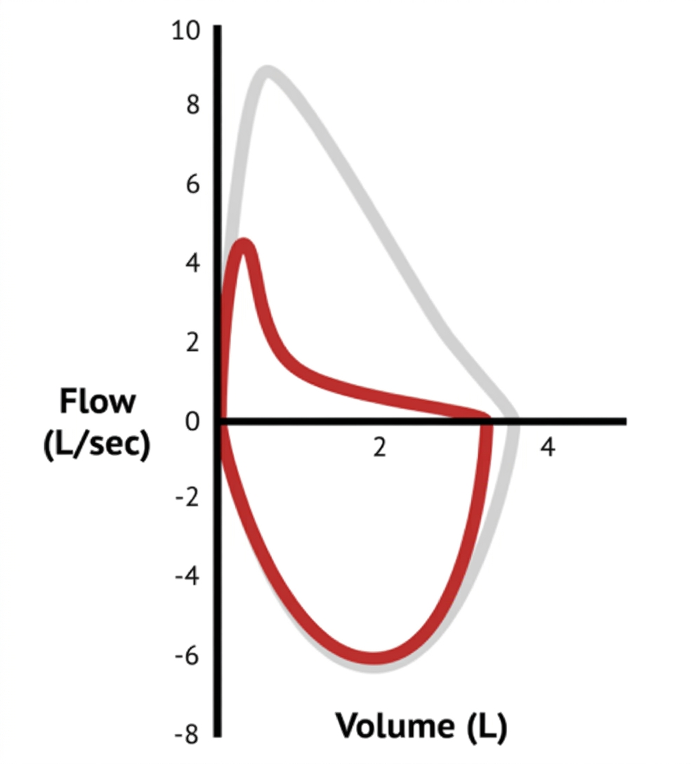
The measured spirometric parameters are recorded below:
- Baseline $FEV_1$: $1.20\text{ L}$ ($58\%$ of predicted)
- Baseline $FVC$: $2.10\text{ L}$ ($84\%$ of predicted)
- Baseline $FEV_1/FVC$ ratio: $0.57$ (Absolute lower limit of normal: $0.80$)
- Post-bronchodilator $FEV_1$: $1.45\text{ L}$ ($70\%$ of predicted)
- Post-bronchodilator $FVC$: $2.18\text{ L}$ ($87\%$ of predicted)
Questions
- Describe the key graphical and numerical characteristics of this flow-volume loop and determine the primary ventilatory defect.
- State the international consensus (ATS/ERS) diagnostic criteria for defining a clinically significant Bronchodilator Reversibility (BDR) in pediatric patients.
- Calculate the absolute change and the percentage change in $FEV_1$ relative to baseline for this patient, and state whether the test confirms bronchodilator responsiveness.
- Detail the standard preparation instructions regarding withholding times for short-acting beta-agonists (SABA) and long-acting beta-agonists (LABA) prior to diagnostic spirometry.
Answer
- Graphical Interpretation & Ventilatory Defect:
- Graphical Pattern: Concave upward ("scooped-out") appearance of the descending expiratory limb of the maximal expiratory flow-volume curve, indicating reduced instantaneous flows at low lung volumes ($FEF_{25-75\%}$).
- Numerical Pattern: Reduced $FEV_1$ ($<80\%$ predicted), normal $FVC$ ($>80\%$ predicted), and a depressed $FEV_1/FVC$ ratio ($0.57$, significantly below LLN of $0.80$ or $z\text{-score} < -1.64$).
- Primary Defect: Moderate Obstructive Ventilatory Defect (consistent with active bronchial asthma).
- ATS/ERS Diagnostic Criteria for BDR:
- In children, an increase in $FEV_1$ (and/or $FVC$) of $>12\%$ relative to the baseline value (or $>10\%$ of predicted $FEV_1$) and an absolute volume increase of $\ge 200\text{ mL}$ indicates significant positive bronchodilator reversibility.
- Mathematical Calculation of Reversibility:
$$\begin{aligned}
\text{Absolute Change in } FEV_1 &= \text{Post-}FEV_1 - \text{Pre-}FEV_1 \\
&= 1.45\text{ L} - 1.20\text{ L} = \mathbf{0.25\text{ L}} \quad (\mathbf{250\text{ mL}}) \\[1em]
\text{Percentage Change from Baseline} &= \frac{\text{Post-}FEV_1 - \text{Pre-}FEV_1}{\text{Pre-}FEV_1} \times 100 \\
&= \frac{0.25}{1.20} \times 100 = \mathbf{20.83\%}
\end{aligned}
$$
- Conclusion: The test confirms statistically significant and clinically positive bronchodilator reversibility ($20.83\% \text{ increase and } 250\text{ mL volume gain}$, exceeding the $>12\%$ and $\ge 200\text{ mL}$ thresholds).
- Medication Withholding Schedule Prior to Testing:
- Short-Acting Beta-2 Agonists (SABA) (e.g., Salbutamol, Levosalbutamol): Withhold for at least $4\text{ to }6\text{ hours}$.
- Short-Acting Muscarinic Antagonists (SAMA) (e.g., Ipratropium): Withhold for at least $12\text{ hours}$.
- Long-Acting Beta-2 Agonists (LABA) (e.g., Formoterol, Salmeterol): Withhold for at least $24\text{ to }48\text{ hours}$.
- Ultra-Long-Acting Beta-2 Agonists (Ultra-LABA) (e.g., Indacaterol, Vilanterol): Withhold for at least $48\text{ hours}$.
RES039 - Child With Otalgia and Fever
Scenario
A 10-month-old male infant is brought to the pediatric clinic with high fever up to 39.2°C (102.5°F), extreme irritability, sleeplessness, and vigorous left ear tugging for 36 hours following a 4-day prodrome of viral upper respiratory tract infection. Pneumatic otoscopy of the symptomatic left ear is performed.
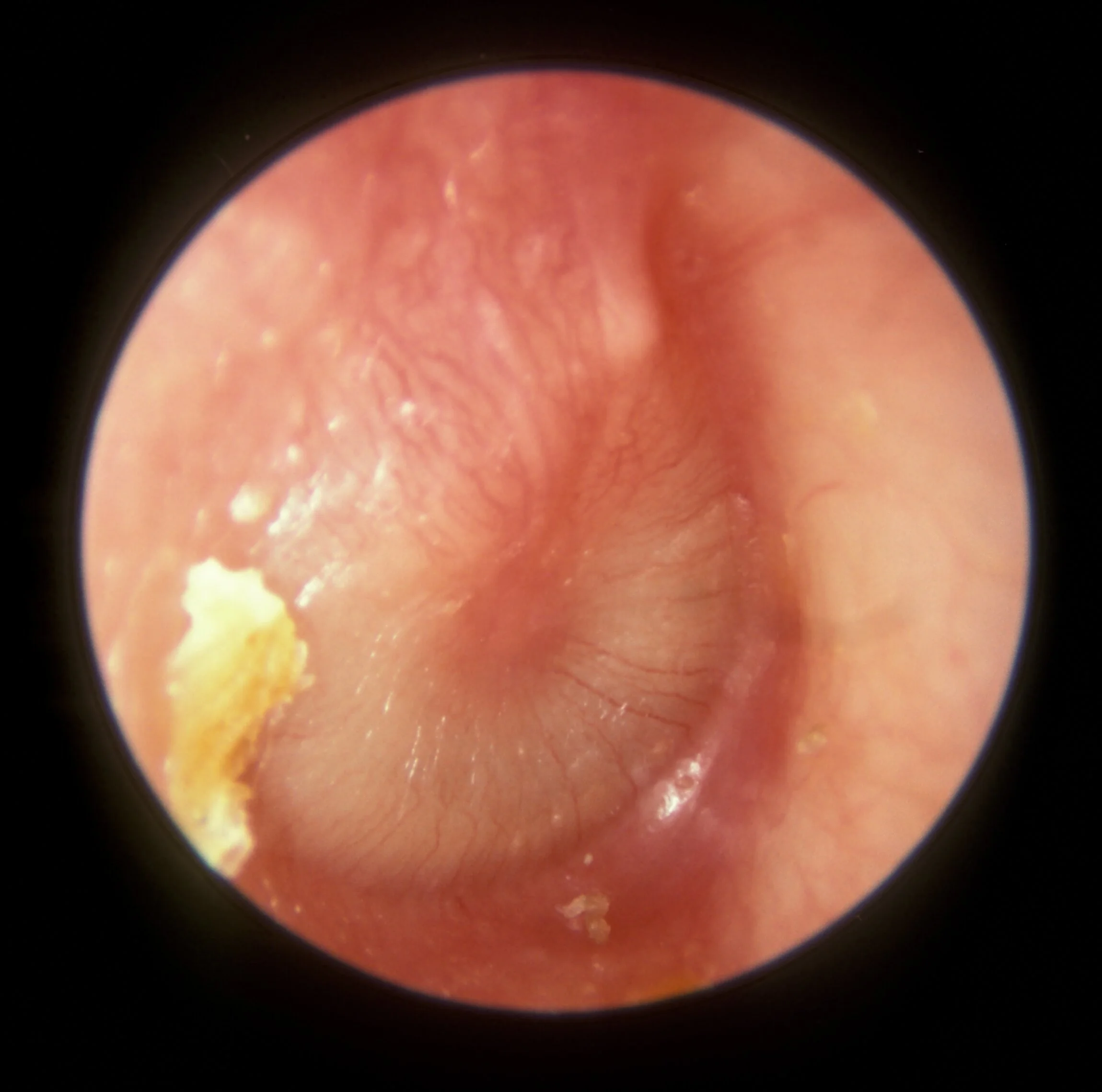
Questions
- Describe the three characteristic otoscopic signs demonstrated on examination and formulate the definitive clinical diagnosis.
- List the three most common bacterial etiologies responsible for this clinical condition in pediatric patients.
- Specify the first-line antibiotic of choice, including exact weight-based daily dosage, dosing interval, and duration. Explain the pharmacological rationale for using high doses.
- Enumerate four evidence-based behavioral and immunization interventions to prevent recurrent episodes in infants.
Answer
- Otoscopic Findings & Diagnosis:
- Findings: Distinct moderate-to-severe outward bulging of the tympanic membrane ("doughnut" appearance), intense diffuse erythema/hyperemia, loss of the normal translucent pearl-gray color and light reflex (cone of light), and obscuration of anatomical ossicular landmarks (handle of malleus).
- Definitive Diagnosis: Acute Otitis Media (AOM), severe left-sided.
- Bacterial Etiologies:
- Streptococcus pneumoniae (predominant pathogen).
- Non-typeable Haemophilus influenzae (NTHi).
- Moraxella catarrhalis.
- First-Line Antibiotic & Pharmacological Rationale:
- Drug & Regimen: Oral Amoxicillin at $80\text{–}90\text{ mg/kg/day}$ administered in 2 divided doses (every 12 hours) for 10 days (in children under 2 years of age).
- Rationale: High-dose amoxicillin achieves high concentrations in middle ear fluid ($>6\ \mu\text{g/mL}$) that exceed the Minimum Inhibitory Concentration (MIC) necessary to saturate and overcome altered penicillin-binding proteins ($PBP\text{-}1a$ and $PBP\text{-}2x$) present in penicillin-intermediate and resistant strains of Streptococcus pneumoniae.
- Preventive Strategies:
- Exclusive breastfeeding for the first 6 months of life.
- Total elimination of environmental tobacco smoke exposure (passive smoking).
- Avoidance of supine bottle feeding (propping bottles) and cessation of pacifier use after 6 months of age.
- Complete vaccination with Pneumococcal Conjugate Vaccine (PCV-10, PCV-13, or PCV-15) and routine annual Influenza vaccination.
RES040 - Worsening Lymphadenopathy During Antimicrobial Therapy
Scenario
A 10-year-old girl with microbiologically confirmed pulmonary tuberculosis and prominent right hilar adenopathy was initiated on standard four-drug anti-tubercular therapy (Isoniazid, Rifampicin, Pyrazinamide, and Ethambutol). She demonstrated initial clinical improvement with resolution of fever, reduction in cough, and weight gain during the first 6 weeks. However, at 8 weeks of therapy, she develops acute bilateral painless cervical lymph node enlargement, low-grade fever, and mild exertional dyspnea without stridor. Direct observation confirms strict treatment adherence. A contrast-enhanced chest CT demonstrates enlargement of subcarinal and paratracheal lymph nodes compressing the right main bronchus, with extensive bilateral necrotic cervical lymphadenitis.
Questions
- Name this clinical phenomenon and define its diagnostic parameters, stating the typical timeframe of onset.
- Outline the immunological mechanism responsible for this clinical presentation.
- List four distinct alternative diagnoses or complications that must be rigorously excluded before confirming this diagnosis.
- Detail the evidence-based management protocol for this patient, addressing anti-tubercular therapy continuation, anti-inflammatory intervention, and surgical indications.
Answer
Phenomenon & Definition:
- Condition: Paradoxical Upgrading Reaction (PUR) / Immune Reconstitution Inflammatory Syndrome (IRIS) in Tuberculosis.
- Definition: The temporary clinical or radiological worsening of pre-existing tuberculous lesions or the unexpected appearance of new tubercular lesions occurring in a patient who previously demonstrated documented clinical improvement on appropriate anti-TB therapy, without evidence of treatment failure or non-adherence.
- Onset Timeline: Typically occurs $2\text{ to }12\text{ weeks}$ (peak incidence at 4 to 8 weeks) after starting effective anti-tubercular treatment.
Immunological Mechanism:
- Rapid and effective bactericidal killing of M. tuberculosis by potent anti-tubercular drugs causes massive lysis of mycobacteria and sudden release of large quantities of mycobacterial antigens (e.g., lipoarabinomannan, antigen 85 complex, cell wall lipids).
- Concurrently, successful anti-tubercular therapy reverses the state of active disease-induced systemic immunosuppression and anergy, leading to a vigorous host cell-mediated immune rebound (delayed-type hypersensitivity).
- Surges of pro-inflammatory Th1 cytokines (interferon-$\gamma$ [IFN-$\gamma$], tumor necrosis factor-$\alpha$ [TNF-$\alpha$], interleukin-2 [IL-2], and interleukin-6 [IL-6]) recruit activated macrophages and lymphocytes, resulting in exuberant granulomatous inflammation, localized vascular permeability, tissue edema, caseation necrosis, and paradoxical swelling of involved lymph nodes.
Alternative Diagnoses & Complications to Rigorously Exclude:
- Secondary Drug-Resistant Tuberculosis: Emergence or unrecognized baseline multi-drug resistant (MDR-TB) or rifampicin-resistant TB (RR-TB); repeat CBNAAT/GeneXpert and culture/drug susceptibility testing (DST) on nodal aspirate.
- Patient Non-Adherence or Drug Malabsorption: Inadequate bioavailability due to irregular drug intake, subtherapeutic dosing, or gastrointestinal malabsorption.
- Secondary Bacterial Superinfection: Acute suppurative pyogenic lymphadenitis caused by Staphylococcus aureus or Streptococcus pyogenes.
- Alternative or Concurrent Malignancy / Systemic Pathology: Hodgkin lymphoma, non-Hodgkin lymphoma, acute leukemia, or atypical mycobacterial infection (NTM).
Evidence-Based Management Protocol:
- Continuation of Anti-Tubercular Therapy:
- Continue the current first-line ATT regimen uninterrupted at full weight-based doses. Reassure the child and parents that paradoxical upgrading reflects immune recovery and bactericidal drug efficacy rather than treatment failure.
- Systemic Anti-Inflammatory Therapy:
- Indicated for moderate-to-severe symptoms, impending airway compromise (tracheobronchial compression), extensive necrotic cervical adenitis, or visual/neurological deficits.
- Administer oral Prednisolone $1\text{ to }2\text{ mg/kg/day}$ (maximum $60\text{ mg/day}$) for $2\text{ to }4\text{ weeks}$, followed by a gradual tapering over the subsequent $2\text{ to }4\text{ weeks}$ based on clinical response (total corticosteroid duration: 4 to 8 weeks).
- Surgical Indications:
- Needle Aspiration: Strongly indicated for large, tense, fluctuant lymph nodes threatening spontaneous breakdown, to alleviate skin tension and prevent persistent scrofuloderma/chronic discharging sinus tracts (prefer needle aspiration using a wide-bore needle entering through normal adjacent skin over incision and drainage).
- Surgical Excision / Decompression: Reserved strictly for critical airway compression refractory to systemic corticosteroids or progressive vascular compression.
- Continuation of Anti-Tubercular Therapy:
RES041 - Febrile Child With Pleurisy
Scenario
A 7-year-old boy presents with high-grade fever, tachypnea, and productive cough for 10 days. Over the last 48 hours, he developed severe left-sided chest pain that worsens with deep inspiration, causing him to tilt his trunk toward the left side. On physical examination, respiratory rate is 46/min, oxygen saturation is 91% on room air, and trachea is shifted to the right. Percussion reveals a stony dull note over the left mid and lower lung zones with absent breath sounds and decreased vocal fremitus. A diagnostic thoracocentesis yields thick, turbid fluid.
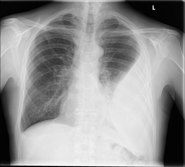 (
Image Source
)
Questions
- Formulate the most specific provisional diagnosis and list the 3 most common bacterial etiologies in this age group.
- Outline the 3 classical stages in the natural progression of this pleural pathology with their temporal characteristics.
- State the pleural fluid biochemical and cytological criteria used to differentiate this condition from an uncomplicated parapneumonic effusion.
- Detail the acute interventional management and first-line empirical antibiotic regimen (with drug name, route, dosage, and duration) for this patient.
Answer
- Provisional Diagnosis & Etiology:
- Diagnosis: Left-sided Empyema Thoracis (Complicated Parapneumonic Effusion).
- Common organisms: Staphylococcus aureus (including CA-MRSA), Streptococcus pneumoniae, and Streptococcus pyogenes (Group A Streptococcus).
- Stages of Progression:
- Stage 1 (Exudative phase, Day 1–3): Thin, sterile fluid with low cellularity; lung remains readily expandable.
- Stage 2 (Fibrinopurulent phase, Day 4–14): Accumulation of polymorphonuclear leukocytes, high fibrin deposition, formation of loculations, septae, and viscous frank pus.
- Stage 3 (Organizing / Fibroblastic phase, > 14 days): Ingrowth of fibroblasts forming a thick, inelastic pleural peel resulting in lung entrapment and thoracic deformity (scoliosis).
- Pleural Fluid Diagnostic Criteria:
- Macroscopic: Frank pus, turbid or cloudy appearance.
- Pleural fluid $\text{pH} < 7.20$.
- Pleural fluid glucose $< 40\text{ mg/dL}$ (or pleural-to-serum ratio $< 0.5$).
- Pleural fluid $\text{LDH} > 1000\text{ IU/L}$ (or pleural-to-serum ratio $> 0.6$).
- Pleural fluid leukocytes $> 50,000/\mu\text{L}$ (predominantly polymorphonuclear neutrophils).
- Positive Gram stain or bacterial culture.
- Management Protocol:
- Thoracic Drainage: Intercostal tube drainage (ICD) with underwater seal (16–20 Fr tube placed in the 4th/5th intercostal space anterior to mid-axillary line) or video-assisted thoracoscopic surgery (VATS) with fibrinolytics (e.g., intrapleural Alteplase/tPA $0.1\text{ mg/kg}$ paired with Dornase alfa).
- Empirical Antibacterial Therapy:
- Intravenous Ceftriaxone $75\text{–}100\text{ mg/kg/day}$ IV once daily (or divided q12h) PLUS
- Intravenous Vancomycin $40\text{–}60\text{ mg/kg/day}$ IV divided q6h or q8h (or Clindamycin $30\text{–}40\text{ mg/kg/day}$ IV divided q8h for MRSA/anaerobic cover).
- Total duration: 2 to 4 weeks (at least 7–10 days post-fever defervescence and tube removal).
More Details
flowchart TD
    A["Parapneumonic Effusion on CXR / USG"] --> B{"Pleural Ultrasound Features"}
    B -->|"Free-flowing, anechoic, pH > 7.20"| C["Conservative: IV Antibiotics"]
    B -->|"Septated, Loculated, Frank Pus, pH < 7.20"| D["Complicated Effusion / Empyema"]
    D --> E["Small-Bore Chest Tube / Pigtail Catheter"]
    E --> F{"Adequate Drainage & Clinical Response within 48–72h?"}
    F -->|"Yes"| G["Continue IV Antibiotics & Wean Drain"]
    F -->|"No / Dense Loculations"| H["Intrapleural Fibrinolysis: tPA + DNase OR Early VATS"]
    H --> I["Decortication if trapped lung persists > 2 weeks"]
RES042 - Office Respiratory Monitoring Tool
Scenario
An 8-year-old girl with moderate persistent asthma is being evaluated during a routine follow-up clinic visit. She has a standing height of 120 cm. As part of her objective pulmonary status assessment and self-management plan, the clinician utilizes the mechanical bedside instrument shown below.
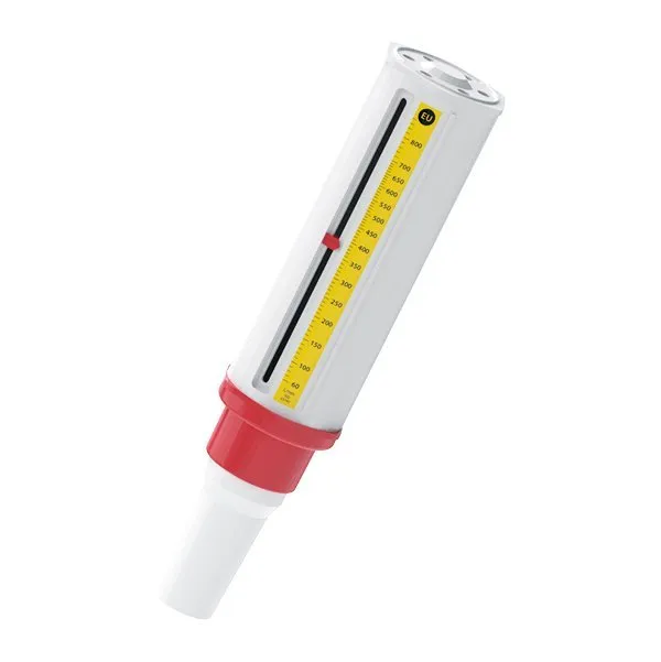
Questions
- Identify the instrument shown and state the specific physiologic parameter it measures.
- Calculate the child's estimated predicted normal value using Godfrey's pediatric mathematical bedside formula.
- List 4 essential standardized steps in patient counseling to ensure accurate measurement using this device.
- Outline the three-tier asthma action plan traffic light zones based on the patient's personal best or predicted value.
Answer
- Identification:
- Device: Wright's Peak Flow Meter (or mini-Wright peak expiratory flow meter).
- Parameter measured: Peak Expiratory Flow Rate (PEFR), expressed in Liters per minute ($\text{L/min}$).
- Mathematical Calculation: $$\begin{aligned} \text{Predicted PEFR (L/min)} &= [\text{Height (in cm)} - 80] \times 5 \\ &= (120 - 80) \times 5 \\ &= 40 \times 5 \\ &= \mathbf{200\text{ L/min}} \quad (180\text{–}220\text{ L/min}) \end{aligned} $$
- Standard Technique Steps:
- Ensure the marker/indicator is reset to zero on the scale.
- Stand up straight (or sit upright consistently) and hold the device horizontally without obstructing the exit slot or marker track with fingers.
- Inhale as deeply and fully as possible (to total lung capacity).
- Place mouthpiece in mouth, seal lips tightly around it (keeping tongue down), and blow out as hard and fast as possible in a single "blast" of expiration.
- Record the best of 3 acceptable maneuvers.
- Action Plan Zones:
- Green Zone (Safe / Controlled): $80\text{–}100\%$ of personal best/predicted ($160\text{–}200\text{ L/min}$) $\rightarrow$ continue baseline controller medications.
- Yellow Zone (Caution / Deterioration): $50\text{–}79\%$ of personal best/predicted ($100\text{–}158\text{ L/min}$) $\rightarrow$ initiate short-acting $\beta_2$-agonist (SABA) rescue therapy and step-up controller as per action plan.
- Red Zone (Medical Alert / Severe Danger): $< 50\%$ of personal best/predicted ($< 100\text{ L/min}$) $\rightarrow$ take immediate bronchodilator therapy and seek emergency medical evaluation.
RES043 - Febrile Toddler With Tachypnea
Scenario
A 22-month-old girl weighing 11 kg is brought to an urban primary health clinic with a history of fever and cough for 3 days. On physical examination, she is alert, interacting with her mother, and drinking fluids normally. She has no stridor, no cyanosis, and no lower chest wall indrawing. Her axillary temperature is 38.6°C, heart rate is 128/min, and respiratory rate measured over a full 60 seconds when calm is 54/min. Auscultation reveals bilateral harsh vesicular breath sounds with scattered right infrascapular crackles.
Questions
- Define the threshold for fast breathing (tachypnea) across the 3 pediatric age brackets per World Health Organization (WHO) / Integrated Management of Neonatal and Childhood Illness (IMNCI) guidelines.
- Classify this child's illness category according to current revised WHO / IMNCI respiratory guidelines.
- List 4 "general danger signs" under IMNCI guidelines that would immediately escalate this classification.
- Specify the first-line oral antibiotic regimen, route, weight-based dosage, frequency, and duration for home-based management of this child.
Answer
- Cut-offs for Fast Breathing (IMNCI / WHO):
- Age $< 2\text{ months}$: $\ge 60\text{ breaths/min}$.
- Age $2\text{ to } 11\text{ months}$: $\ge 50\text{ breaths/min}$.
- Age $12\text{ to } 59\text{ months}$: $\ge 40\text{ breaths/min}$.
- IMNCI Classification:
- Pneumonia (Child has cough or difficulty breathing with fast breathing [$\ge 40\text{/min}$ for a 22-month-old], but without chest indrawing or general danger signs).
- General Danger Signs:
- Inability to drink or breastfeed.
- Vomiting everything ingested.
- History of convulsions during the current illness episode.
- Lethargy or unconsciousness.
- (Additionally: Stridor in a calm child or severe malnutrition indicates immediate hospitalization).
- Antibiotic Treatment:
- First-line: Oral Amoxicillin.
- Dose: High-dose dispersible tablets at $80\text{–}90\text{ mg/kg/day}$ divided into 2 doses (every 12 hours) [Standard WHO alternative: $40\text{–}50\text{ mg/kg/day}$ divided twice daily].
- Calculation for 11 kg child: $11\text{ kg} \times 80\text{ mg/kg/day} = 880\text{ mg/day} \rightarrow 440\text{–}500\text{ mg}$ orally twice daily.
- Duration: 5 days (in non-severe pneumonia, a 3-day course of high-dose amoxicillin is also endorsed in low-risk settings).
RES044 - Recurrent Childhood Expiratory Wheeze
Scenario
A 9-year-old boy presents with a 6-month history of nocturnal dry cough, recurrent chest tightness, and shortness of breath precipitated by running during sports class. He is clinically stable at presentation. A diagnostic pulmonary investigation is conducted in the laboratory, producing a volume-time curve and flow-volume loop with pre- and post-inhalation measurements.
Questions
- Identify the formal diagnostic investigation and describe 3 graphical features on the flow-volume loop indicative of intrathoracic lower airway obstruction.
- State the primary physiological indices evaluated on this report and specify the cut-off defining an obstructive ventilatory defect.
- Formulate the precise spirometric criteria used to establish significant bronchodilator reversibility in children.
- List 4 clinical indications and 2 absolute/relative contraindications for performing this procedure in a pediatric patient.
Answer
- Investigation & Loop Morphology:
- Investigation: Pulmonary Function Testing (PFT) / Spirometry with Pre- and Post-Bronchodilator Flow-Volume Loop.
- Morphologic features of lower airway obstruction:
- "Scooped-out" or concave appearance of the expiratory limb (coving).
- Significant reduction in Peak Expiratory Flow (PEF).
- Normal or preserved inspiratory limb (as obstruction is predominantly intrathoracic).
- Physiological Indices & Obstructive Defect:
- Primary parameters: Forced Expiratory Volume in 1 second ($\text{FEV}_1$), Forced Vital Capacity ($\text{FVC}$), $\text{FEV}_1/\text{FVC}$ ratio, and Forced Expiratory Flow at 25–75% of vital capacity ($\text{FEF}_{25-75\%}$).
- Criteria for obstruction: Reduced $\text{FEV}_1/\text{FVC}$ ratio below the lower limit of normal (LLN, typically $< 0.80\text{–}0.85$ or $< 80\text{–}85\%$ in children).
- Bronchodilator Reversibility Criteria:
- An increase in $\text{FEV}_1$ of $\ge 12\%$ from baseline AND $\ge 200\text{ mL}$ absolute increase (or $\ge 12\%$ increase in predicted $\text{FEV}_1$) 15 minutes following inhalation of $400\ \mu\text{g}$ of Salbutamol via metered-dose inhaler with spacer.
- Indications and Contraindications:
- Indications:
- Differentiation between obstructive and restrictive lung disorders.
- Diagnosis and monitoring of bronchial asthma.
- Pre-operative pulmonary risk assessment.
- Longitudinal monitoring of chronic lung diseases (e.g., cystic fibrosis, bronchopulmonary dysplasia).
- Contraindications:
- Recent pneumothorax (within 4–6 weeks).
- Recent thoracic, abdominal, or ophthalmic surgery.
- Hemodynamically unstable congenital heart disease or thoracic aneurysm.
- Active hemoptysis of unknown etiology.
- Indications:
RES045 - Hypoxemic Full-Term Neonatal Distress
Scenario
A term male neonate born at 39 weeks via emergency cesarean section for thick meconium-stained amniotic fluid develops severe tachypnea, grunting, and central cyanosis within 2 hours of birth. At 12 hours of life, he is intubated and mechanically ventilated on pressure-control mode. Ventilator settings: $\text{FiO}_2 = 1.0\ (100\%)$, Peak Inspiratory Pressure $(\text{PIP}) = 32\text{ cm H}_2\text{O}$, Positive End-Expiratory Pressure $(\text{PEEP}) = 6\text{ cm H}_2\text{O}$, Mean Airway Pressure $(\text{MAP}) = 15\text{ cm H}_2\text{O}$. Simultaneous pulse oximetry demonstrates pre-ductal (right hand) $\text{SpO}_2$ of 94% and post-ductal (left foot) $\text{SpO}_2$ of 81%. Arterial blood gas (ABG) from the right radial artery shows: $\text{pH } 7.22$, $\text{PaCO}_2\ 48\text{ mm Hg}$, $\text{PaO}_2\ 50\text{ mm Hg}$, $\text{HCO}_3^-\ 19\text{ mEq/L}$, Base Deficit $-7.8\text{ mEq/L}$.
Questions
- Interpret the underlying pathophysiologic mechanism explaining the differential pulse oximeter saturation between the pre-ductal and post-ductal sites.
- Calculate the baby's Oxygenation Index (OI) and state the severity category.
- What is the clinical implication of this calculated OI regarding targeted advanced pulmonary vasodilator and extracorporeal therapies?
- Outline the pharmacotherapy protocol for this condition, detailing the initial drug of choice, route, starting dose, and second-line systemic vasodilators.
Answer
- Mechanism of Pre- and Post-Ductal Gradient:
- Suprasystemic pulmonary vascular resistance causes right-to-left shunting of deoxygenated blood across a patent ductus arteriosus (PDA) into the descending aorta.
- The right subclavian artery (pre-ductal) receives better-oxygenated blood from the ascending aorta, whereas post-ductal lower limbs receive desaturated blood diluted by the right-to-left ductal shunt (difference $\ge 10\%$ is diagnostic of PPHN).
- Mathematical Calculation:
$$\begin{aligned}
\text{Oxygenation Index (OI)} &= \frac{\text{MAP } (\text{cm H}_2\text{O}) \times \text{FiO}_2\ (\%)}{\text{PaO}_2\ (\text{mm Hg})} \\
&= \frac{15 \times 100}{50} \\
&= \mathbf{30} \quad (\text{Normal: } < 10)
\end{aligned}
$$
- Severity: Severe hypoxemic respiratory failure / pulmonary hypertension ($\text{OI } 25\text{–}40$).
- Clinical Implications of OI:
- $\text{OI} \ge 20\text{–}25$: Absolute indication for initiating Inhaled Nitric Oxide (iNO) therapy.
- $\text{OI} \ge 40$ (or $\text{OI} > 35$ despite optimal iNO and HFOV for 4–6 hours): Meets standard entry criteria for Extracorporeal Membrane Oxygenation (ECMO).
- Pharmacotherapy Protocol:
- First-line Inhaled Vasodilator: Inhaled Nitric Oxide (iNO) started at $20\text{ ppm}$ via continuous inspiratory limb delivery.
- Adjunctive Inodilator / Inotropes (to maintain systemic BP > pulmonary artery pressure):
- Dopamine ($5\text{–}10\ \mu\text{g/kg/min}$) and/or Epinephrine ($0.05\text{–}0.2\ \mu\text{g/kg/min}$) to keep mean systemic arterial pressure above pulmonary pressures.
- Milrinone: Loading dose $50\ \mu\text{g/kg}$ over 30 min (often omitted if hypotensive) followed by maintenance infusion of $0.33\text{–}0.75\ \mu\text{g/kg/min}$ IV (improves RV performance and pulmonary vascular resistance).
- Oral / IV Phosphodiesterase-5 (PDE-5) Inhibitor (where iNO is unavailable/refractory):
- Sildenafil: $1\text{–}2\text{ mg/kg/dose}$ orally/NG q6h (or IV Sildenafil loading $0.4\text{ mg/kg}$ over 3 hours, then $1.6\text{ mg/kg/day}$ continuous infusion).
More Details
flowchart TD
    A["Term/Near-term Infant with Severe Hypoxemia"] --> B["Demonstrate Pre-Post Ductal SpO2 Gradient > 10% or Echocardiography confirmation"]
    B --> C["Optimize Ventilation: HFOV / Surfactant if parenchymal disease"]
    C --> D["Calculate Oxygenation Index: MAP x FiO2 / PaO2"]
    D -->|"OI 15 - 25"| E["Optimize Sedation, Correct Acidosis, Normotension: Dopamine / Milrinone"]
    D -->|"OI >= 25"| F["Initiate Inhaled Nitric Oxide at 20 ppm"]
    F --> G{"Assess Response at 30-60 mins: PaO2 rise > 20 mmHg or SpO2 rise"}
    G -->|"Responder"| H["Gradually wean iNO by 5 ppm every 4 hours down to 5 ppm"]
    G -->|"Non-responder / OI >= 40"| I["Consider Cannulation for ECMO"]
RES046 - Infant Paroxysmal Cough and Cyanosis
Scenario
A 5-month-old female infant is brought to the pediatric emergency department with a 2-week history of worsening respiratory symptoms. Her illness began with mild rhinorrhea, low-grade fever, and an intermittent dry cough. Over the past 4 days, she has developed episodic spasms of staccato coughing occurring in paroxysms, frequently accompanied by facial cyanosis, post-tussive emesis, and inability to feed. The mother reports no inspiratory whoop. The infant received only birth vaccines (BCG, OPV-0, Hepatitis B) and missed all subsequent immunizations. On examination: Heart rate 168/min, Respiratory rate 52/min, $\text{SpO}_2$ 96% on room air between spasms (drops to 78% during spasms), capillary refill time 2 seconds. Systemic examination reveals petechiae over the forehead and bilateral subconjunctival hemorrhages. Chest auscultation reveals clear breath sounds between paroxysms without wheeze or crackles. Complete blood count reveals: Hemoglobin 11.2 g/dL, Total leukocyte count $48,000/\mu\text{L}$ with 78% mature lymphocytes.
Questions
- State the most probable diagnosis and explain why an inspiratory "whoop" is absent in this patient.
- List two gold-standard laboratory investigations to confirm this diagnosis and state the preferred specimen collection technique.
- Calculate the Absolute Lymphocyte Count (ALC) and explain the cellular mechanism responsible for the marked lymphocytosis.
- Specify the first-line antimicrobial therapy with precise dosing, route, and duration, along with the post-exposure prophylaxis recommendation for household contacts.
Answer
- Primary Diagnosis & Absence of Whoop:
- Diagnosis: Pertussis (Whooping Cough) caused by Bordetella pertussis.
- Absence of Whoop: Young infants under 6 months of age have immature respiratory mechanics, smaller airway calibers, and insufficient inspiratory force to generate the classic loud whoop; instead, they typically present with paroxysmal staccato cough, apnea, post-tussive cyanosis, bradycardia, or seizures.
- Confirmatory Investigations & Specimen Collection:
- Polymerase Chain Reaction (PCR): Real-time PCR targeting insertion sequences (e.g., IS481 and IS1001), highly sensitive within the first 3–4 weeks of illness.
- Bacterial Culture: Inoculation on selective enriched media (Regan-Lowe charcoal agar or Bordet-Gengou medium).
- Specimen Technique: Dacron or calcium alginate swab (not cotton) inserted deeply into the posterior nasopharynx and held for several seconds, or nasopharyngeal aspirate.
- Absolute Lymphocyte Count Calculation & Pathophysiology:
$$\begin{aligned}
\text{Absolute Lymphocyte Count (ALC)} &= \text{Total Leukocyte Count} \times \left(\frac{\text{Lymphocyte } \%}{100}\right) \\
&= 48{,}000/\mu\text{L} \times 0.78 \\
&= \mathbf{37{,}440/\mu\text{L}} \quad (\text{Normal infant range: } 3{,}000 - 9{,}500/\mu\text{L})
\end{aligned}
$$
- Pathophysiological Mechanism: Pertussis toxin (PT) mediates ADP-ribosylation of the inhibitory alpha subunit ($G_{\alpha i}$) of heterotrimeric G proteins. This disrupts chemokine receptor signaling, selectively blocking the extravasation and homing of mature T and B lymphocytes from the vascular compartment into lymph nodes, trapping them in circulation (lymphocytosis-promoting factor effect).
- Antimicrobial Therapy and Prophylaxis:
- First-Line Drug: Azithromycin oral suspension, $10\text{ mg/kg/day}$ once daily for 5 days (preferred in infants $<1$ month and older infants due to minimal risk of infantile hypertrophic pyloric stenosis compared to erythromycin).
- Alternative: Clarithromycin oral suspension, $15\text{ mg/kg/day}$ divided every 12 hours ($7.5\text{ mg/kg/dose}$) for 7 days.
- Household Post-Exposure Prophylaxis (PEP): Administer oral Azithromycin at the same therapeutic dosing to all close household contacts regardless of their age or immunization status, initiated within 21 days of cough onset in the index patient.
More Details
RES047 - Post-Infectious Cystic Chest Radiograph
Scenario
A 22-month-old boy is recovering from severe community-acquired pneumonia requiring 7 days of intravenous antibiotics and high-flow nasal cannula oxygenation. Over the past 48 hours, his fever has subsided, tachypnea has improved, and he is weaning successfully from supplemental oxygen. A routine follow-up frontal chest radiograph is obtained to assess resolution of the extensive consolidation.
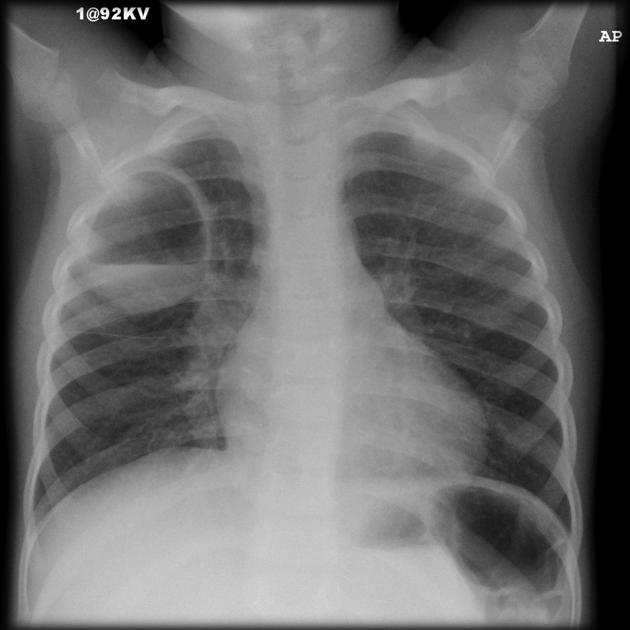 (
Image Source
)
Questions
- Identify the radiological abnormality shown on the chest radiograph.
- Name the two most common bacterial pathogens associated with the formation of these parenchymal lesions in young children.
- Describe the underlying pathophysiological mechanism responsible for lesion formation.
- Outline the definitive management strategy for uncomplicated lesions versus the specific indications requiring percutaneous or surgical decompression.
Answer
- Radiological Abnormality:
- Pulmonary pneumatoceles (post-infectious subpleural / intraparenchymal thin-walled, air-filled cystic lesions).
- Common Bacterial Pathogens:
- Staphylococcus aureus (particularly Methicillin-Resistant S. aureus [MRSA] producing Panton-Valentine leukocidin [PVL]).
- Klebsiella pneumoniae (or Streptococcus pneumoniae, particularly serotypes 3 and 19A).
- Pathophysiology:
- Necrotizing inflammation causes focal bronchiolar wall ulceration, mucosal necrosis, and peribronchial edema.
- Intraluminal fibrinous exudates create a one-way "check-valve" or ball-valve obstruction.
- Air enters the distal parenchymal spaces during inspiration but cannot escape during expiration, resulting in alveolar distension, rupture of interalveolar septa, and progressive cystic ballooning in the subpleural or pulmonary interstitial plane.
- Management Strategy:
- Uncomplicated Lesions: Conservative, expectant management with continuation of culture-directed systemic antibiotics. The vast majority (>85%) spontaneously regress and completely resolve over weeks to months (typically 2–12 months) without long-term parenchymal distortion.
- Indications for Intervention:
- "Tension" pneumatocele causing compression of adjacent normal lung, mediastinal shift, and acute cardiorespiratory compromise.
- Rupture into the pleural space producing tension pneumothorax or pyopneumothorax.
- Secondary bacterial superinfection leading to intra-cystic abscess formation unresponsive to medical therapy.
- Interventions: Image-guided percutaneous pigtail catheter decompression/drainage or surgical resection (lobectomy/wedge resection).
RES048 - Subcutaneous Crepitus and Mediastinal Radiolucency
Scenario
A 7-year-old girl with a history of acute severe asthma is intubated and mechanically ventilated in the pediatric intensive care unit due to hypercapnic respiratory failure. On day 2 of invasive ventilation with high peak inspiratory pressures ($34\text{ cmH}_2\text{O}$), the bedside nurse notes sudden swelling over the anterior neck and supraclavicular regions. Palpation reveals extensive crackling subcutaneous crepitus. Cardiovascular auscultation reveals distant heart sounds with a distinct raspy, clicking sound synchronous with the heartbeat. An urgent portable chest radiograph is performed.
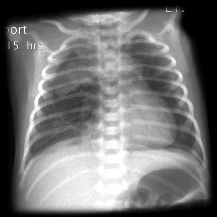 (
Image Source
)
Questions
- Identify the primary diagnostic radiological finding demonstrated on the chest radiograph.
- Name the pathognomonic physical auscultatory sign described and detail its clinical features.
- Explain the pathophysiological sequence (Macklin effect) leading to this condition.
- List two iatrogenic ICU procedures/interventions that precipitate this condition, and outline the initial therapeutic management.
Answer
- Radiological Diagnosis:
- Pneumomediastinum (mediastinal emphysema) with extensive subcutaneous emphysema (along with typical signs such as the continuous diaphragm sign, thymic sail sign / angel wing sign, or ring-around-the-artery sign).
- Auscultatory Physical Sign:
- Hamman sign (Hamman's crunch / click / murmur):
- A raspy, crunching, scratchy, or clicking sound heard over the precordium during cardiac systole (and occasionally diastole), synchronous with the heartbeats.
- It is accentuated during expiration and best appreciated when the patient is placed in the left lateral decubitus position.
- Hamman sign (Hamman's crunch / click / murmur):
- Pathophysiology (Macklin Effect):
- High intra-alveolar pressure (due to positive pressure ventilation and air trapping in severe asthma) creates a steep pressure gradient between the marginal alveoli and the lung interstitium.
- Terminal alveoli rupture, allowing gas to escape into the pulmonary interstitium.
- Centripetal gas dissection occurs along the peribronchovascular sheaths toward the pulmonary hila.
- Air enters the mediastinum (pneumomediastinum) and subsequently tracks along contiguous fascial planes into the neck, retropharyngeal space, and subcutaneous tissue of the chest wall.
- Iatrogenic Causes and Initial Management:
- Iatrogenic Causes:
- High peak inspiratory pressures / barotrauma from invasive mechanical ventilation.
- Endotracheal intubation (tracheobronchial laceration or perforation during difficult intubation).
- Central venous catheterization (subclavian or internal jugular vein puncture traversing the pleura/fascia).
- Diagnostic/therapeutic rigid bronchoscopy.
- Management Strategy:
- Ventilator Optimization: Implement lung-protective ventilation (reduce tidal volume to $4-6\text{ mL/kg}$, reduce peak inspiratory pressure, reduce PEEP, allow permissive hypercapnia) to prevent further alveolar rupture.
- Conservative / Supportive Care: Most cases are self-limiting; high-concentration supplemental oxygen (nitrogen washout therapy) accelerates reabsorption of encapsulated mediastinal gas.
- Decompression: Needle aspiration or mediastinotomy/blow-hole subcutaneous incisions are reserved only for rare "tension pneumomediastinum" causing vena caval compression and obstructive shock.
- Iatrogenic Causes:
RES049 - Postural Drainage Positioning for Atelectasis
Scenario
A 6-year-old child with cystic fibrosis is admitted to the pediatric pulmonary ward with acute pulmonary exacerbation, copious viscid secretions, and persistent segmental atelectasis. High-resolution computed tomography and chest radiography confirm subsegmental collapse and thick mucous impaction in the right middle lobe and the lingular segments of the left upper lobe. The pediatric team prescribes targeted airway clearance therapy including postural drainage.
Questions
- Describe the precise patient posture, body rotation, and tilt elevation required for postural drainage of the lateral and medial segments of the right middle lobe.
- Describe the precise patient posture, body rotation, and tilt elevation required for postural drainage of the superior and inferior segments of the left lingula.
- State four absolute or relative contraindications to placing a pediatric patient in the Trendelenburg (head-down) position during chest physiotherapy.
- Enumerate four active or passive chest physiotherapy techniques that should be applied concurrently with postural drainage positioning.
Answer
- Postural Drainage Position for Right Middle Lobe:
- Posture: Supine position with the body turned one-quarter turn to the left.
- Support: Right side supported with pillows or wedges placed from the shoulder to the hip to maintain the quarter-turn tilt.
- Table / Bed Elevation: Foot end of the bed elevated by 14 inches ($35\text{ cm}$), corresponding to a $15^\circ$ to $20^\circ$ Trendelenburg angle.
- Postural Drainage Position for Left Lingular Segments:
- Posture: Supine position with the body turned one-quarter turn to the right.
- Support: Left side supported with pillows or wedges from the left shoulder to the hip to maintain the quarter-turn tilt.
- Table / Bed Elevation: Foot end of the bed elevated by 14 inches ($35\text{ cm}$), corresponding to a $15^\circ$ to $20^\circ$ Trendelenburg angle.
- Contraindications to Trendelenburg / Head-Down Positioning:
- Increased intracranial pressure (e.g., neurotrauma, hydrocephalus, meningitis, intracranial hemorrhage).
- Severe gastroesophageal reflux disease (GERD) with high risk of aspiration.
- Hemodynamic instability, uncontrolled hypertension, or decompensated congestive heart failure.
- Acute massive hemoptysis.
- Marked abdominal distension, bowel obstruction, or recent postprandial state ($<1-2\text{ hours}$ after feeding).
- Concurrent Adjunctive Techniques:
- Manual percussion (chest wall cupping using a cupped hand or soft silicone palm cup).
- Chest wall vibration and rib springing during exhalation.
- Active Cycle of Breathing Techniques (ACBT) / forced expiratory technique (huffing).
- Oscillating positive expiratory pressure (PEP) therapy (e.g., Flutter, Acapella, or high-frequency chest wall oscillation vest).
RES050 - Prolonged Childhood Fever and Cough
Scenario
A 4-year-old boy is brought to the pediatric outpatient department with a history of low-to-moderate grade continuous fever for the past 3 weeks and an unremitting cough that began 2 weeks ago. His mother notes that he has lost weight over the past 3 months and has lost his appetite. His paternal grandmother, who resided in the same household, was diagnosed with pulmonary tuberculosis and died one month prior. Physical examination reveals: Weight $11.5\text{ kg}$ ($<3\text{rd}$ percentile for age and sex, severe underweight), Height $96\text{ cm}$ (10th percentile), mild pallor, no peripheral lymphadenopathy, no clubbing. Respiratory examination reveals normal vesicular breath sounds without adventitious sounds. Abdominal examination is unremarkable.
Questions
- Define "Presumptive Pediatric Tuberculosis" according to the National Tuberculosis Elimination Program (NTEP) diagnostic guidelines.
- Outline the diagnostic algorithm and specific biological specimens recommended for microbiological confirmation via upfront molecular testing in this child.
- State the cut-off values for a positive Tuberculin Skin Test (Mantoux test) in immunocompetent children versus children with Severe Acute Malnutrition (SAM) or HIV infection.
- Specify the current NTEP drug regimen, daily weight-based drug doses, and duration for treating drug-susceptible pediatric pulmonary tuberculosis in this patient.
Answer
- NTEP Definition of Presumptive Pediatric Tuberculosis:
- A child presenting with any of the following clinical features:
- Persistent fever for $>2$ weeks without a known etiology.
- Unremitting cough for $>2$ weeks.
- Weight loss ($>5\%$ decrease in body weight within the past 3 months) or failure to gain weight over the past 3 months despite nutritional support.
- History of close contact with an adult or adolescent index case of pulmonary tuberculosis within the preceding 2 years.
- Unexplained, persistent cervical/peripheral lymphadenopathy or non-resolving chest radiograph findings.
- A child presenting with any of the following clinical features:
- Specimen Collection and Diagnostic Cascade:
- Specimens: Two early morning gastric aspirates/lavages (collected after $\ge 4$ hours of overnight fasting) or induced sputum specimens (obtained using $3\%$ hypertonic saline nebulization).
- First-Line Diagnostic Test: Upfront Cartridge-Based Nucleic Acid Amplification Test (CBNAAT / GeneXpert MTB/RIF) or Truenat to simultaneously detect Mycobacterium tuberculosis complex and rifampicin resistance.
- Complementary Tests: Digital chest radiograph (PA and lateral views) and Tuberculin Skin Test (TST) using 2 tuberculin units (TU) of PPD RT-23.
- Mantoux Test (TST) Cut-Off Criteria:
- General Immunocompetent Children: Induration $\ge 10\text{ mm}$ transverse diameter at 48–72 hours is considered positive.
- Immunocompromised / SAM / HIV Children: Induration $\ge 5\text{ mm}$ transverse diameter at 48–72 hours is considered positive (due to suppressed cell-mediated cutaneous hypersensitivity).
- NTEP Drug Regimen and Weight-Based Dosing:
- Regimen: Daily fixed-dose combination (FDC) regimen administered orally:
- Intensive Phase (IP - 2 Months): 4 drugs — Isoniazid (H), Rifampicin (R), Pyrazinamide (Z), Ethambutol (E) [2 HRZE].
- Continuation Phase (CP - 4 Months): 3 drugs — Isoniazid (H), Rifampicin (R), Ethambutol (E) [4 HRE].
- Weight-Based Daily Dosages:
- Isoniazid (H): $10\text{ mg/kg/day}$ (range $7-15\text{ mg/kg/day}$)
- Rifampicin (R): $15\text{ mg/kg/day}$ (range $10-20\text{ mg/kg/day}$)
- Pyrazinamide (Z): $35\text{ mg/kg/day}$ (range $30-40\text{ mg/kg/day}$)
- Ethambutol (E): $20\text{ mg/kg/day}$ (range $15-25\text{ mg/kg/day}$)
- Formulation for $11.5\text{ kg}$ (10–15 kg weight band):
- IP: 3 tablets daily of pediatric dispersible FDC (HRZ $50/75/150\text{ mg}$) plus 3 tablets daily of Ethambutol ($100\text{ mg}$).
- CP: 3 tablets daily of pediatric dispersible FDC (HRE $50/75/100\text{ mg}$).
- Regimen: Daily fixed-dose combination (FDC) regimen administered orally:
More Details
- Presumptive symptoms persistent $>2$ weeks,
- Consistent chest radiograph abnormalities (mediastinal/hilar lymphadenopathy, miliary pattern, parenchymal consolidation), and
- Evidence of TB infection (positive Mantoux test) and/or documented close household contact with an adult index case. Response to a broad-spectrum antibiotic course (excluding fluoroquinolones, macrolides, or aminoglycosides) must show complete absence of clinical improvement before initiating empiric anti-tubercular therapy.
RES051 - Supraglottic Airway Device Clinical Evaluation
Scenario
A 3-year-old child weighing 14 kg with persistent middle lobe atelectasis is scheduled for diagnostic flexible fiberoptic bronchoscopy and bronchoalveolar lavage under procedural sedation. The pediatric anesthesiologist selects the supraglottic device shown in the photograph to secure the airway while maintaining continuous gastric venting.
Questions
- Identify the specific medical device shown in the exhibit and describe four distinct anatomical design components.
- State two primary clinical advantages of this specific device over a first-generation Classic Laryngeal Mask Airway (cLMA).
- State the correct device size and the maximum permissible cuff inflation volume for this 14 kg child.
- List two absolute or relative contraindications to the elective placement of this device.
Answer
- Device Identification and Anatomical Components:
- Device: ProSeal Laryngeal Mask Airway (PLMA / ProSeal LMA).
- Design Components:
- Dual-lumen shaft: Parallel respiratory tube (for ventilation) and drain tube (for gastric decompression).
- Posterior dorsal cuff: Additional dorsal inflatable cuff providing a superior mucosal seal against the hypopharynx.
- Integral bite block: Built-in silicone block preventing tube occlusion from accidental biting.
- Gastric drain orifice: Distal tip opening positioned at the upper esophageal sphincter to vent gas or pass an orogastric tube.
- Pilot balloon with one-way check valve: For monitoring and titrating cuff inflation pressure.
- Clinical Advantages over Classic LMA:
- Higher Oropharyngeal Seal Pressure: Achieves an airtight seal up to $30\text{ cmH}_2\text{O}$ (compared to $\approx 20\text{ cmH}_2\text{O}$ with cLMA), permitting positive pressure ventilation without gastric insufflation.
- Gastric Decompression and Aspiration Protection: Drain tube allows insertion of a gastric decompression tube and vents regurgitated stomach contents away from the laryngeal inlet.
- Absence of Aperture Bars: Permits unimpeded passage of a flexible fiberoptic bronchoscope.
- Device Sizing and Cuff Volume: $$\begin{aligned} \text{Device Size} &= \mathbf{\text{Size 2}} \quad (10 - 20\text{ kg}) \\ \text{Max Inflation Volume} &= \mathbf{10\text{ mL}} \quad (\text{Target cuff pressure } < 60\text{ cmH}_2\text{O}) \\ \text{Max Orogastric Tube Size} &= \mathbf{10\text{ Fr}} \end{aligned} $$
- Contraindications:
- Full stomach, active gastroesophageal reflux, or delayed gastric emptying in an emergent, non-fasted setting where rapid-sequence endotracheal intubation is indicated.
- Severe pharyngeal pathology, retropharyngeal abscess, or mass occluding the supraglottic space.
- Severely reduced pulmonary compliance or high airway resistance requiring peak inspiratory pressures $>30\text{ cmH}_2\text{O}$.
- Severe trismus, inter-incisor opening $<1.5\text{ cm}$, or restricted mouth opening.
RES052 - Ventilated Child Acid Base Derangement
Scenario
A 4-year-old child weighing 16 kg with severe necrotizing pneumonia is on mechanical ventilation in the pediatric intensive care unit. Current ventilator settings: Volume-Controlled Continuous Mandatory Ventilation (VC-CMV), PEEP: $6\text{ cmH}_2\text{O}$, Tidal Volume ($V_T$): $96\text{ mL}$ ($6\text{ mL/kg}$), Respiratory Rate: $20\text{ breaths/min}$, $\text{FiO}_2$: $0.50$, Inspiratory Time ($T_i$): $0.75\text{ s}$.
Arterial blood gas (ABG) reveals:
- $\text{pH}: 7.18$
- $\text{PaCO}_2: 68\text{ mmHg}$
- $\text{PaO}_2: 82\text{ mmHg}$
- $\text{HCO}_3^-: 25.2\text{ mEq/L}$
- $\text{Base Excess (BE)}: -3.2\text{ mEq/L}$
Questions
- Formulate the primary acid-base diagnosis.
- Determine whether this is a simple or mixed disorder by calculating the expected bicarbonate compensation.
- Calculate the target ventilator respiratory rate required to normalize the $\text{PaCO}_2$ to $40\text{ mmHg}$, assuming constant dead-space fraction and carbon dioxide production.
- Outline four bedside interventions (ventilator and airway circuit adjustments) to correct this condition.
Answer
- Primary Acid-Base Diagnosis:
- Acute, uncompensated (or minimally compensated) respiratory acidosis with acceptable oxygenation ($\text{PaO}_2/\text{FiO}_2 = 164$, mild-to-moderate ARDS).
- Physiological Compensation and Disorder Classification:
$$\begin{aligned}
\Delta\text{PaCO}_2 &= 68 - 40 = 28\text{ mmHg} \\
\text{Expected }\Delta\text{HCO}_3^-\text{ (Acute)} &= 0.1 \times \Delta\text{PaCO}_2 = 0.1 \times 28 = 2.8\text{ mEq/L} \\
\text{Expected }[\text{HCO}_3^-] &= 24 + 2.8 = \mathbf{26.8\text{ mEq/L}} \quad (\text{Range: } 25.0 - 28.0\text{ mEq/L})
\end{aligned}
$$
- The measured $[\text{HCO}_3^-]$ of $25.2\text{ mEq/L}$ matches the acute physiological compensation buffer range. This represents a simple, uncompensated/acutely buffered respiratory acidosis without an underlying independent metabolic derangement.
- Target Respiratory Rate Calculation: $$\begin{aligned} \text{Target RR} &= \frac{\text{Current RR} \times \text{Current }\text{PaCO}_2}{\text{Target }\text{PaCO}_2} \\ &= \frac{20 \times 68}{40} \\ &= \mathbf{34\text{ breaths/min}} \end{aligned} $$
- Bedside Interventions to Alleviate Hypercapnia:
- Increase Alveolar Minute Ventilation: Increase ventilator respiratory rate toward $30 - 34\text{ breaths/min}$ (monitoring for intrinsic PEEP / air-trapping) or titrate tidal volume to $7 - 8\text{ mL/kg}$ ($112 - 128\text{ mL}$) if driving pressure ($P_{\text{plat}} - \text{PEEP}$) remains $<15\text{ cmH}_2\text{O}$.
- Clear Mechanical Airway Occlusion: Perform endotracheal suctioning to remove endobronchial mucus plugs and clear secretions.
- Reduce Circuit Dead Space: Remove unneeded heat and moisture exchangers (HME), extenders, or stopcocks between the Y-piece and endotracheal tube; transition to an active heated-wire humidifier.
- Treat Reversible Bronchospasm: Administer inhaled bronchodilators (e.g., salbutamol nebulization) if expiratory wheezing or expiratory flow limitation with delayed return to baseline is visible on flow-time scalar.
RES053 - Arterial Blood Gas Parameter Interpretation
Scenario
A 2-month-old infant weighing 4.2 kg is on mechanical ventilation following surgical closure of an omphalocele. Routine arterial blood gas (ABG) sampled from an indwelling radial arterial line yields the following parameters:
- $\text{pH}: 7.56$
- $\text{PaCO}_2: 23.7\text{ mmHg}$
- $\text{PaO}_2: 157\text{ mmHg}$
- $\text{HCO}_3^-: 21.0\text{ mmol/L}$
- $\text{SaO}_2: 100\%$
Co-oximetry confirms a total hemoglobin ($\text{Hb}$) concentration of $10.0\text{ g/dL}$.
Questions
- Formulate the primary acid-base and oxygenation diagnosis.
- Calculate the total arterial oxygen content ($\text{CaO}_2$) in $\text{mL O}_2/\text{dL}$ (or $\text{vol}\%$) showing complete working.
- Calculate the expected serum bicarbonate for this acute derangement to verify if metabolic compensation has occurred.
- State three adverse pathophysiological consequences of this acute alkalemic state.
- List two ventilator modifications to normalize the arterial blood gas.
Answer
- Primary Diagnosis:
- Acute uncompensated respiratory alkalosis (alveolar hyperventilation) with hyperoxia.
- Arterial Oxygen Content Calculation: $$\begin{aligned} \text{CaO}_2 &= \left(1.34 \times \text{Hb} \times \frac{\text{SaO}_2}{100}\right) + (0.003 \times \text{PaO}_2) \\ &= (1.34 \times 10.0 \times 1.0) + (0.003 \times 157) \\ &= 13.40 + 0.471 \\ &= \mathbf{13.87\text{ mL O}_2/\text{dL}} \quad (\text{Normal reference: } 16 - 20\text{ mL/dL for age}) \end{aligned} $$
- Expected Serum Bicarbonate Calculation:
$$\begin{aligned}
\Delta\text{PaCO}_2 &= 40 - 23.7 = 16.3\text{ mmHg} \\
\text{Expected }\Delta\text{HCO}_3^-\text{ (Acute)} &= 0.2 \times \Delta\text{PaCO}_2 = 0.2 \times 16.3 = 3.26\text{ mmol/L} \\
\text{Expected }[\text{HCO}_3^-] &= 24 - 3.26 = \mathbf{20.74\text{ mmol/L}} \quad (\text{Range: } 19.5 - 22.0\text{ mmol/L})
\end{aligned}
$$
- The observed $[\text{HCO}_3^-]$ of $21.0\text{ mmol/L}$ matches the acute cellular buffering response perfectly, confirming a pure acute respiratory alkalosis without renal compensation.
- Pathophysiological Consequences:
- Cerebral Hypoperfusion: Hypocapnia induces potent cerebral arteriolar vasoconstriction, decreasing cerebral blood flow and increasing ischemic risk.
- Left Shift of Oxyhemoglobin Dissociation Curve: Increased affinity of hemoglobin for oxygen (Bohr effect), impairing peripheral tissue oxygen offloading.
- Electrolyte Derangements and Arrhythmias: Hypocalcemia (increased protein binding of ionized calcium causing neuromuscular irritability, tetany, or seizure activity) and intracellular shift of potassium causing hypokalemia and cardiac dysrhythmias.
- Ventilator Modifications:
- Decrease Minute Ventilation: Reduce the set respiratory rate or reduce peak inspiratory pressure (PIP) / tidal volume ($V_T$).
- Wean $\text{FiO}_2$: Titrate inspired oxygen downward immediately to target normoxia ($\text{SpO}_2\ 94 - 98\%$, $\text{PaO}_2\ 60 - 80\text{ mmHg}$).
RES054 - Pediatric Spirometry Pattern Clinical Assessment
Scenario
An 11-year-old boy presenting with progressive dry cough and exertional breathlessness for 6 months undergoes computerized spirometry. Clinical examination reveals bilateral basilar end-inspiratory fine crackles and grade 2 digital clubbing.
| Parameter | Measured Value | Predicted Value | Percent Predicted (%) |
|---|---|---|---|
| FVC | $1.13\text{ L}$ | $2.07\text{ L}$ | $54.6\%$ |
| FEV$_1$ | $1.08\text{ L}$ | $1.87\text{ L}$ | $57.7\%$ |
| FEV$_1$/FVC Ratio | $95.6\%$ | $87.0\%$ | $109.9\%$ |
| FEF$_{25-75\%}$ | $1.89\text{ L/s}$ | $2.26\text{ L/s}$ | $83.6\%$ |
Questions
- Interpret the spirometry findings and grade the severity of the physiological impairment.
- Identify the single confirmatory static lung volume test and cut-off value required to formally establish this diagnosis.
- Explain how Diffusing Capacity of the Lung for Carbon Monoxide ($\text{DLCO}$) differentiates intrinsic parenchymal disorders from extrinsic disorders displaying this spirometry pattern.
- List four pediatric causes of this ventilatory defect grouped by anatomical compartment (two intrinsic, two extrinsic).
Answer
- Spirometry Interpretation and Severity:
- Pattern: Restrictive ventilatory defect (proportionate reduction in both FVC and FEV$_1$ with a normal or supranormal $\text{FEV}_1/\text{FVC ratio} > 85\%$).
- Severity: Moderate to Moderately Severe impairment ($\text{FVC } 50 - 59\%$ of predicted, per ATS/ERS guidelines).
- Confirmatory Test and Diagnostic Criterion:
- Plethysmography / Helium Dilution Static Lung Volumes: Demonstration of a reduced Total Lung Capacity (TLC).
- Criterion: $\text{TLC} < 80\%$ of predicted (or below the 5th percentile / lower limit of normal, $Z\text{-score} < -1.645$).
- Role of DLCO in Differentiation:
- Intrinsic Parenchymal Disease: $\text{DLCO}$ is reduced ($<80\%$ predicted or $
- Extrinsic Chest Wall / Neuromuscular Disease: $\text{DLCO}$ is typically normal when corrected for alveolar volume ($\text{DLCO/VA}$), because the alveolar-capillary membrane itself remains structurally intact despite chest wall restriction or muscular weakness.
- Intrinsic Parenchymal Disease: $\text{DLCO}$ is reduced ($<80\%$ predicted or $
- Etiologies by Compartment:
- Intrinsic Parenchymal Disorders (any two):
- Childhood Interstitial Lung Disease (chILD) / Idiopathic pulmonary fibrosis / Desquamative interstitial pneumonitis.
- Pulmonary alveolar proteinosis / Inherited surfactant metabolism dysfunction (mutations in SFTPB, SFTPC, ABCA3).
- Hypersensitivity pneumonitis (extrinsic allergic alveolitis, e.g., bird fancier's lung, farmer's lung).
- Post-infectious or organizing pneumonia / Obliterative bronchiolitis with parenchymal scarring.
- Chemotherapy-induced pulmonary fibrosis (e.g., Bleomycin, Busulfan) or thoracic radiation pneumonitis.
- Extrinsic Disorders (Chest Wall / Neuromuscular / Pleural / Extrathoracic) (any two):
- Chest Wall Deformities: Severe kyphoscoliosis, pectus excavatum, asphyxiating thoracic dystrophy (Jeune syndrome).
- Neuromuscular Disorders: Duchenne muscular dystrophy, spinal muscular atrophy (SMA), myasthenia gravis, Guillain-Barré syndrome, bilateral diaphragmatic paralysis (phrenic nerve palsy).
- Pleural Pathology: Large or organizing pleural effusion, fibrothorax / chronic pleural thickening.
- Extrathoracic / Abdominal Restriction: Massive ascites, gross hepatosplenomegaly, morbid pediatric obesity (obesity hypoventilation syndrome).
- Intrinsic Parenchymal Disorders (any two):
RES055 - National Respiratory Protocol Assessment
Scenario
A 6-year-old child living with HIV (CD4 count: 240 cells/$\mu\text{L}$, on abacavir + lamivudine + dolutegravir) is brought to the pediatric chest clinic with a 3-week history of low-grade fever, unremitting cough, and failure to gain weight. Physical examination reveals bilateral cervical lymphadenopathy and reduced air entry over the right middle zone. Gastric aspirates are obtained for evaluation under the National Tuberculosis Elimination Program (NTEP / formerly RNTCP).
Questions
- Differentiate between "New" and "Previously Treated" tuberculosis patient categorizations under national programmatic guidelines.
- State the laboratory criterion for defining a smear-positive pulmonary tuberculosis case under revised programmatic guidelines compared to the historical definition.
- Outline the clinical screening definition of a "presumptive tuberculosis" case in a child living with HIV.
- Detail the therapeutic considerations and drug modifications required for managing tuberculosis-HIV co-infection in this patient.
- State the revised consensus definition of Extensively Drug-Resistant Tuberculosis (XDR-TB).
Answer
- Categorization of Tuberculosis Cases:
- New Case: A patient who has never had treatment for tuberculosis or has taken anti-tubercular drugs for less than 1 month (28 days).
- Previously Treated Case: A patient who has received 1 month (28 days) or more of anti-tubercular drugs in the past. Subcategories include:
- Relapse: Declared cured/completed treatment in the past and subsequently diagnosed with bacteriologically positive or clinically active TB.
- Treatment after Failure: Received treatment and remained smear/culture-positive at the end of the treatment course.
- Treatment after Loss to Follow-up (formerly Default): Interrupted treatment for 1 month or more consecutively and returns with active TB.
- Definition of Smear-Positive Case:
- Revised NTEP / RNTCP Criterion: Presence of at least one sputum or gastric aspirate sample positive for Acid-Fast Bacilli (AFB) on Ziehl-Neelsen (ZN) staining or LED fluorescence microscopy (a single positive smear suffices).
- Historical Criterion: Required at least two positive smear microscopy examinations, or one positive smear combined with chest radiographic abnormalities suggestive of active TB and physician authorization.
- Presumptive TB Definition in Children Living with HIV:
- Presence of any one of the following symptoms:
- Current cough of any duration (even < 2 weeks)
- Persistent fever of any duration
- Poor weight gain / faltering growth / severe acute malnutrition (SAM)
- History of contact with a confirmed or suspected active pulmonary TB case
- Presence of any one of the following symptoms:
- Tuberculosis-HIV Co-infection Management:
- Anti-Tubercular Therapy (ATT): Daily weight-banded fixed-dose combinations (FDC) consisting of 2 months of HRZE (Isoniazid, Rifampicin, Pyrazinamide, Ethambutol) followed by 4 months of HRE (Isoniazid, Rifampicin, Ethambutol). Rifampicin is maintained throughout.
- Timing of Antiretroviral Therapy (ART): Continue ART or initiate within 2 weeks of starting ATT (once tolerated) to reduce mortality, except in TB meningitis where ART is delayed 4–8 weeks to avoid life-threatening intracranial Immune Reconstitution Inflammatory Syndrome (IRIS).
- Pharmacokinetic Interactions (Dolutegravir): Rifampicin is a potent inducer of CYP3A4 and UGT1A1, markedly lowering Dolutegravir (DTG) trough concentrations. The DTG dose must be given twice daily (an extra supplemental dose 12 hours after the standard daily dose) throughout ATT and continued for 2 weeks after Rifampicin cessation.
- Prophylaxis: Co-trimoxazole Preventive Therapy (CPT) must be continued/initiated simultaneously.
- Revised Definition of XDR-TB (WHO / NTEP):
- Infection with a Mycobacterium tuberculosis strain meeting the criteria for Multidrug-Resistant / Rifampicin-Resistant TB (MDR/RR-TB) that is:
- Resistant to any fluoroquinolone (Levofloxacin or Moxifloxacin), AND
- Resistant to at least one additional Group A drug (Bedaquiline or Linezolid).
- Infection with a Mycobacterium tuberculosis strain meeting the criteria for Multidrug-Resistant / Rifampicin-Resistant TB (MDR/RR-TB) that is:
RES056 - Recurrent Pediatric Pulmonary Infections
Scenario
A 2-year-old boy presents with a history of recurrent right lower lobe respiratory tract infections since 6 months of age. On examination, he has mild tachypnea, reduced breath sounds over the right hemithorax, and dextroposition of the apex beat. Initial chest radiography demonstrated reduced right hemithoracic volume, rightward shift of the cardiomediastinal structures, and an abnormal curvilinear vascular shadow descending along the right cardiac border toward the diaphragm. Imaging performed is shown here.
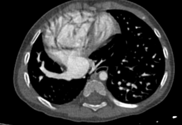 (
Image Source
)
Questions
- Identify the imaging modality shown and describe the two classic imaging abnormalities pathognomonic of this condition.
- What is the definitive clinical diagnosis, and what is its alternative anatomical designation?
- List three associated cardiopulmonary or vascular anomalies characteristically seen with this syndrome.
- What are the indications for surgical intervention, and what surgical technique is employed for definitive repair?
Answer
- Imaging Modality & Pathognomonic Features:
- Contrast-enhanced computed tomography (CECT) of the chest / CT Pulmonary Angiography.
- Right lung hypoplasia with ipsilateral mediastinal and cardiac displacement.
- Anomalous right pulmonary vein draining into the systemic venous circulation, specifically the inferior vena cava (IVC) or the cavoatrial junction below or at the diaphragm (resembling a curved Turkish sword/scimitar).
- Diagnosis & Classification:
- Scimitar syndrome.
- Alternative anatomical name: Congenital pulmonary venolobar syndrome (or Halasz syndrome).
- Associated Cardiopulmonary Anomalies:
- Atrial septal defect (ASD), most frequently secundum type (present in 40–80%).
- Systemic arterial supply to the hypoplastic right lower lobe arising directly from the descending thoracic or abdominal aorta (pulmonary sequestration).
- Hypoplasia or absence of the right pulmonary artery.
- Bronchial branching anomalies (e.g., right bronchial isomerism, tracheal bronchus, diverticula).
- Right diaphragmatic hernia or eventration.
- Indications and Surgical Management:
- Indications: Hemodynamically significant left-to-right shunt ($Q_p:Q_s > 1.5:1$), recurrent pulmonary infections, severe pulmonary hypertension, or symptomatic heart failure.
- Definitive Procedure: Surgical redirection/rerouting of the anomalous pulmonary venous drainage into the left atrium using an intra-atrial baffle (or direct venous reimplantation into the posterior wall of the left atrium) $\pm$ patch closure of any accompanying ASD, along with transcatheter coil embolization or surgical ligation of systemic collateral feeder arteries supplying the sequestered lung segment.
More Details
- Infantile form: Presents early (<1 year of age) with tachypnea, heart failure, and severe pulmonary hypertension. Often associated with large aortopulmonary collateral arteries and intrinsic congenital heart disease; carries a guarded prognosis.
- Childhood/Adult form: Typically diagnosed incidentally or during evaluation for recurrent pneumonias; shunting is modest, pulmonary arterial pressures are usually normal or mildly elevated, and prognosis after repair is excellent.
RES057 - Classification of Severe Mycobacterial Disease
Scenario
A 4-year-old girl weighing 12 kg is brought to the pediatric clinic with persistent non-remitting cough, low-grade evening fever, and anorexia for 8 weeks. Physical examination reveals moderate pallor, marked wasting, and generalized lymphadenopathy. A diagnosis of tuberculosis is confirmed. You are evaluating the child to categorize disease severity to determine the exact therapeutic regimen and duration according to National Tuberculosis Elimination Program (NTEP) and World Health Organization (WHO) pediatric guidelines.
Questions
- Define the criteria used to classify pediatric tuberculosis as "Severe Disease".
- Name four specific extrapulmonary manifestations that automatically fulfill the criteria for severe TB.
- Formulate the recommended pharmacotherapeutic regimen (drugs, phases, and duration) for:
- Severe non-CNS, non-osteoarticular tuberculosis.
- Central nervous system (CNS) tuberculosis / Tuberculous meningitis.
- Specify the recommended adjuvant corticosteroid drug, exact dose, and duration indicated in severe pediatric CNS or pericardial tuberculosis.
Answer
- Criteria for Severe Pediatric TB:
- Presence of cavitary lesions on chest radiography.
- Bilateral or extensive parenchymal involvement (e.g., lobar consolidation, bronchopneumonia pattern, or mass-like consolidation).
- Severe acute malnutrition (SAM) with TB (weight-for-height $<-3\text{ SD}$).
- Advanced HIV infection / severe immunosuppression.
- Acute respiratory distress or hypoxemia requiring supplemental oxygen.
- Presence of severe extrapulmonary manifestations.
- Severe Extrapulmonary Forms:
- Tuberculous meningitis (TBM) and tuberculoma.
- Miliary / Disseminated tuberculosis.
- Spinal tuberculosis (Pott's spine) with or without neurological deficit.
- Tuberculous pericarditis.
- Abdominal tuberculosis with intestinal obstruction, perforation, or peritonitis.
- Treatment Regimens:
- Severe non-CNS, non-osteoarticular TB:
- Intensive Phase (2 months): 4 drugs—Isoniazid (H), Rifampicin (R), Pyrazinamide (Z), and Ethambutol (E) daily.
- Continuation Phase (4 months): 3 drugs—Isoniazid (H), Rifampicin (R), and Ethambutol (E) daily (total 6 months).
- CNS Tuberculosis (TBM) / Spinal TB:
- Intensive Phase (2 months): 4 drugs—HRZE daily.
- Continuation Phase (10 months): 2 to 3 drugs—HRE or HR daily (total duration: 12 months).
- Weight-band dosing for pediatric fixed-dose combinations (FDCs):
- Rifampicin: $15\text{ mg/kg/day}$ (range $10\text{--}20$).
- Isoniazid: $10\text{ mg/kg/day}$ (range $7\text{--}15$).
- Pyrazinamide: $35\text{ mg/kg/day}$ (range $30\text{--}40$).
- Ethambutol: $20\text{ mg/kg/day}$ (range $15\text{--}25$).
- Severe non-CNS, non-osteoarticular TB:
- Adjuvant Corticosteroid Therapy:
- Drug: Prednisolone (oral) or Dexamethasone (IV/oral).
- Dose: Prednisolone $2\text{ mg/kg/day}$ orally (or Dexamethasone $0.4\text{ mg/kg/day}$ IV).
- Duration: Full dose for 4 weeks, followed by a gradual taper over the next 2 to 4 weeks (total duration: 6 to 8 weeks).
RES058 - Spirometric Airway Obstruction Analysis
Scenario
During a pediatric pulmonology clinic, maximal expiratory and inspiratory flow-volume loops (MEFV / MIFV) are obtained from three patients presenting with persistent noisy breathing and airway obstruction. The three distinct configurations are marked as Loop A, Loop B, and Loop C.
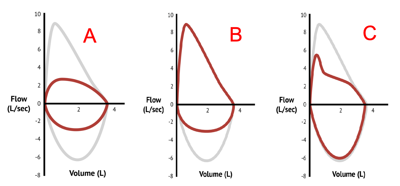
Questions
- Determine the exact anatomical site and dynamic classification of airway obstruction demonstrated in Loops A, B, and C.
- Name the physiological flow ratio measured at mid-vital capacity and state its expected diagnostic value in Loop B versus Loop C.
- List two representative pediatric etiologies for the pattern depicted in Loop A and two for Loop B.
- Describe the physical mechanism (transmural pressure dynamics) explaining why Loop B selectively truncates inspiratory flow while preserving expiratory flow.
Answer
- Classification of Obstruction:
- Loop A: Fixed airway obstruction (intra- or extrathoracic).
- Loop B: Variable extrathoracic upper airway obstruction.
- Loop C: Variable intrathoracic central airway obstruction.
- Mid-Vital Capacity Ratio:
- Ratio: Mid-vital capacity flow ratio = $\frac{FEF_{50\%}}{FIF_{50\%}}$ (Forced Expiratory Flow at 50% VC / Forced Inspiratory Flow at 50% VC). $$\begin{aligned} \text{Normal Value} &\approx 0.8 - 1.0 \\ \text{Loop B (Variable Extrathoracic)} &> \mathbf{1.0} \quad (\text{often } > 2.0\text{ due to marked drop in } FIF_{50\%}) \\ \text{Loop C (Variable Intrathoracic)} &< \mathbf{0.3 - 0.5} \quad (\text{due to selective reduction in } FEF_{50\%}) \end{aligned} $$
- Representative Pediatric Etiologies:
- Loop A (Fixed): Concentric subglottic stenosis (post-intubation/congenital), complete vascular ring (double aortic arch), tracheal web, fixed tracheal foreign body.
- Loop B (Variable Extrathoracic): Laryngomalacia, bilateral or unilateral vocal cord paralysis/paresis, subglottic hemangioma, pharyngeal hypotonia / obstructive sleep apnea.
- Transmural Pressure Dynamics for Loop B:
- During inspiration, atmospheric pressure surrounding the extrathoracic trachea/larynx exceeds the subatmospheric (negative) intraluminal pressure generated to draw air into the lungs. This negative transmural pressure gradient causes dynamic inward collapse of compliant or mobile extrathoracic airway segments, severely restricting inspiratory flow (flattened MIFV loop).
- During expiration, positive intraluminal pressure relative to surrounding atmospheric pressure splints the compliant extrathoracic airway open, allowing normal or near-normal expiratory flow (preserved MEFV loop).
RES059 - Persistent Neonatal Inspiratory Noise
Scenario
A 6-week-old term infant is evaluated for chronic noisy breathing that commenced at 10 days of age. The mother reports a high-pitched sound that worsens during crying, agitation, and when placed in the supine position, but substantially improves when the baby is placed prone. The infant is exclusively breastfed, has no history of cyanosis, fever, or feeding interruptions, and is tracking normally along the 50th percentile for weight and length.
Questions
- State the most probable diagnosis and the gold-standard investigation required for definitive confirmation.
- Enumerate three classic dynamic endoscopic findings observed during this procedure.
- Categorize congenital or early infantile stridor into persistent versus episodic/recurrent causes, giving three examples of each.
- State three absolute clinical indications for surgical intervention in this condition.
Answer
- Most Probable Diagnosis & Confirmation:
- Congenital laryngomalacia.
- Gold Standard: Awake flexible fiberoptic nasopharyngolaryngoscopy (revealing dynamic supraglottic collapse during spontaneous tidal breathing).
- Classic Endoscopic Findings:
- Inward prolapse of redundant, edematous arytenoid mucosa and cuneiform cartilages into the laryngeal inlet on inspiration.
- Foreshortened, tight aryepiglottic folds tethering the epiglottis posteriorly.
- Inward collapsing, tubular, elongated "omega-shaped" ($\Omega$) epiglottis retroflexing over the airway during inspiration.
- Differential Diagnosis by Temporal Pattern:
- Persistent Stridor:
- Congenital subglottic stenosis.
- Complete vascular ring (e.g., double aortic arch, pulmonary artery sling).
- Congenital laryngeal web or atresia.
- Vocal fold paralysis (bilateral or unilateral).
- Congenital laryngeal cyst (vallecular/saccular cyst).
- Episodic / Recurrent Stridor:
- Recurrent respiratory papillomatosis (RRP).
- Proliferating subglottic hemangioma (symptoms expand over months and fluctuate with crying/URTI).
- GERD-induced secondary laryngospasm.
- Viral laryngotracheobronchitis (croup).
- Spasmodic croup.
- Persistent Stridor:
- Absolute Indications for Supraglottoplasty:
- Severe airway compromise: Resting stridor with severe sternal/intercostal retractions, cyanotic spells, or intermittent obstructive apnea.
- Significant feeding difficulty resulting in failure to thrive (FTT) / weight faltering.
- Recurrent aspiration pneumonia or witnessed life-threatening aspiration events.
- Development of cor pulmonale or pectus excavatum deformity.
RES060 - National Tuberculosis Notification Protocol
Scenario
A 10-year-old child attending a private pediatric health facility is diagnosed with pulmonary tuberculosis based on a positive sputum Cartridge-Based Nucleic Acid Amplification Test (CBNAAT / GeneXpert MTB/RIF). As the consultant pediatrician, you are initiating anti-tubercular therapy and fulfilling mandatory statutory public health notification obligations under the Government of India's National Tuberculosis Elimination Program (NTEP).
Questions
- Name the official national web-based surveillance portal for case registration, and explain the linguistic etymology of its title.
- State the statutory penal provisions applicable to medical practitioners and healthcare establishments under Indian law for failure to notify a tuberculosis case.
- Name the direct benefit transfer (DBT) scheme linked to this portal that provides nutritional support, specifying the financial entitlement and disbursement mechanism.
- Identify the specific digital identification code generated by this portal for lifelong individual patient tracking.
Answer
- National Portal & Etymology:
- Portal Name: Ni-kshay (NIKSHAY).
- Etymology: Derived from the Hindi/Sanskrit words:
- "Ni" = 'Without' / 'To end' / 'Eradicate'.
- "Kshay" = 'Tuberculosis' / 'Phthisis' / 'Decay'.
- Meaning: "Eradication of Tuberculosis".
- Statutory Penal Provisions (as per Bharatiya Nyaya Sanhita, BNS 2023 / IPC):
- Under the Ministry of Health and Family Welfare Gazette Notification and the Clinical Establishments Act, failure to notify a tuberculosis case attracts penal liability:
- Section 271, BNS 2023 (formerly Section 269, IPC): Negligent act likely to spread infection of disease dangerous to life (imprisonment up to 6 months, or fine, or both).
- Section 272, BNS 2023 (formerly Section 270, IPC): Malignant act likely to spread infection of disease dangerous to life (imprisonment up to 2 years, or fine, or both).
- Section 223, BNS 2023 (formerly Section 188, IPC): Disobedience to an order duly promulgated by a public servant.
- Healthcare facilities also face administrative penalties including suspension or cancellation of their operating registration/license.
- Nutritional Support Scheme:
- Scheme Name: Ni-kshay Poshan Yojana (NPY).
- Entitlement: ₹500 per month (revised to ₹1,000 per month under updated NTEP guidelines) for each completed month of anti-tubercular therapy.
- Mechanism: Direct Benefit Transfer (DBT) directly into the beneficiary's (or parent/guardian's) Aadhaar-seeded operational bank account via Public Financial Management System (PFMS).
- Unique Digital Identifier:
- Ni-kshay ID (a unique, computerized multi-digit episode/patient identification number).
RES061 - Asymptomatic Household Tuberculosis Contact
Scenario
A 3-year-old boy weighing 14 kg is brought to the pediatric outpatient department by his mother. The child's father was diagnosed one week ago with microbiologically confirmed, rifampicin-sensitive pulmonary tuberculosis (sputum smear 2+ acid-fast bacilli). The child has no history of fever, cough, anorexia, or weight loss. On physical examination, the child is active, well-nourished (weight-for-age at 50th percentile), afebrile, and respiratory system examination is unremarkable with vesicular breath sounds bilaterally. A screening chest radiograph reveals normal lung parenchyma without hilar lymphadenopathy or consolidation.
Questions
- What initial steps must be systematically performed before prescribing TB preventive therapy (TPT) to this child?
- Detail the recommended TB preventive therapy regimens (drugs, dosing frequency, and duration) approved for drug-susceptible tuberculosis child contacts.
- Calculate the precise daily dose of Isoniazid for this 14 kg child if selecting the 6-month monotherapy (6H) regimen, and state the maximum allowable pediatric daily dose.
- What are the key clinical monitoring parameters and indications to discontinue TPT during follow-up visits?
Answer
- Prerequisites Prior to Initiating TPT:
- Rule out active tuberculosis disease: Detailed screening for symptoms (current cough, fever, poor weight gain, lethargy) and abnormal physical findings.
- Radiographic screening: Normal chest radiograph (anteroposterior/posteroanterior) confirming absence of intrathoracic lymphadenopathy or parenchymal lesions.
- Determine drug susceptibility profile of the source contact (confirm rifampicin/isoniazid susceptibility).
- Note: Tuberculin Skin Test (TST) / Interferon Gamma Release Assay (IGRA) is not mandatory to initiate TPT in asymptomatic household contacts aged <5 years under national guidelines (NTEP/WHO).
- Recommended TPT Regimens for Drug-Susceptible TB Contacts:
- 6H (Isoniazid Monotherapy): Daily Isoniazid for 6 months (180 doses).
- 3HR (Isoniazid + Rifampicin): Daily combined Isoniazid and Rifampicin for 3 months (84 doses) in children of all ages.
- 3HP (Isoniazid + Rifapentine): Once-weekly Isoniazid plus Rifapentine for 12 weeks (12 doses) in children aged $\ge 2$ years.
- Dose Calculation (6H Regimen): $$\begin{aligned} \text{Daily Isoniazid Dose} &= 10\text{ mg/kg/day (Range: } 7\text{--}15\text{ mg/kg/day for children } <10\text{ years)} \\ &= 10\text{ mg/kg} \times 14\text{ kg} \\ &= \mathbf{140\text{ mg/day orally once daily}} \quad (\text{Maximum daily dose: } \mathbf{300\text{ mg/day}}) \end{aligned} $$
- Monitoring Parameters and Indications for Discontinuation:
- Monthly assessment for breakthrough signs/symptoms of active TB (cough, unremitting fever, weight loss).
- Assessment of drug adverse events: Jaundice, recurrent vomiting, persistent nausea, epigastric pain, or unexplained lethargy (hepatotoxicity).
- Monitor adherence via pill counts or blister pack review.
- Discontinuation criteria: Development of active TB disease (transition immediately to full multi-drug antitubercular therapy) or clinical jaundice/transaminases $>5 \times$ upper limit of normal (ULN) asymptomatic or $>3 \times$ ULN symptomatic.
More Details
RES062 - Pediatric Antitubercular Therapy Default
Scenario
A 4-year-old child weighing 12 kg with microbiologically confirmed pulmonary tuberculosis was initiated on first-line antitubercular therapy (ATT) comprising daily 4-drug fixed-dose combinations (HRZE: Isoniazid, Rifampicin, Pyrazinamide, Ethambutol). After completing 3 weeks of the intensive phase, the family relocated to their native village, and all medications were completely stopped. The child returns to the clinic after an interruption period.
Questions
- Define the algorithmic management of treatment interruption if the duration of missed doses is less than 4 weeks (<28 consecutive days).
- Outline the clinical and microbiological management protocol if the treatment interruption is 4 weeks or longer ($\ge 28$ consecutive days).
- Which specific investigations must be ordered when restarting therapy following an interruption of $\ge 4$ weeks?
- How is the treatment duration adjusted when an interruption occurs during the continuation phase?
Answer
- Management of Treatment Interruption <4 Weeks (<28 Days):
- Resume the previous regimen immediately without restarting from day zero.
- Compensate for the missed doses by extending the duration of the current phase so that the child receives the complete planned number of doses (e.g., full 56 daily doses for 8-week intensive phase).
- Management of Treatment Interruption $\ge 4$ Weeks ($\ge 28$ Days):
- Perform comprehensive clinical reassessment and collect fresh respiratory/gastric aspirate specimens for microbiological evaluation.
- If the child is clinically symptomatic or smear/culture/NAAT positive: Restart the full treatment regimen from the beginning (Day 1 of Intensive Phase: 2 months HRZE).
- If the child is entirely asymptomatic and smear/culture negative: Base continuation on total cumulative therapy received prior to default and multidisciplinary team consensus.
- Mandatory Investigations on Interruption $\ge 4$ Weeks:
- Rapid molecular drug susceptibility testing: Cartridge Based Nucleic Acid Amplification Test (CBNAAT / GeneXpert MTB/RIF or Truenat) to evaluate for emergent rifampicin resistance.
- Repeat sputum / gastric aspirate / induced sputum for acid-fast bacilli (AFB) smear microscopy and liquid culture (MGIT).
- Second-line Line Probe Assay (LPA) or phenotypic drug susceptibility testing if rifampicin resistance is detected.
- Repeat baseline chest radiograph to assess radiological progression or cavitation.
- Baseline liver function tests (AST, ALT, total and direct bilirubin).
- Adjustment for Interruption during Continuation Phase:
- If interruption is <4 weeks: Resume and extend the continuation phase to complete the total required daily doses (112 doses in a 16-week continuation phase).
- If interruption is $\ge 4$ weeks and cumulative doses completed are $<80\%$ of total planned treatment: Restart the complete treatment course (both intensive and continuation phases) while awaiting drug susceptibility test results.
- If $\ge 80\%$ of doses were completed and the patient remains asymptomatic with negative smear/culture: Treatment may be closed as "Treatment Completed" under close post-treatment observation.
RES063 - Pediatric Tuberculosis Outcome Classifications
Scenario
A district pediatric tuberculosis coordinator is auditing treatment completion and success metrics for a cohort of 120 pediatric patients registered under the National Tuberculosis Elimination Program (NTEP) on the Nikshay portal. Standardized programmatic outcome definitions must be assigned at the end of the treatment course.
Questions
- State the exact operational definition of "Cured" and differentiate it from "Treatment Completed".
- Define "Treatment Failed" under national tuberculosis elimination guidelines.
- Define the outcomes "Died" and "Lost to Follow-up" in programmatic surveillance.
- Define the outcome "Treatment Regimen Changed" and list two clinical scenarios that mandate this entry.
Answer
- "Cured" vs "Treatment Completed":
- Cured: An initially microbiologically confirmed pulmonary TB patient who was smear- or culture-negative in the last month of treatment and on at least one previous occasion during treatment.
- Treatment Completed: A patient who completed treatment as recommended by the national guidelines, without evidence of failure, but who lacks documented negative smear or culture results in the last month of treatment and on at least one previous occasion (either because tests were not done or in clinically diagnosed/extrapulmonary TB cases).
- "Treatment Failed":
- A TB patient whose biological specimen (smear or culture) is positive at the end of treatment (or at month 5 or later during therapy); OR
- A patient whose treatment regimen needed to be terminated or permanently changed to a new regimen due to lack of clinical improvement, clinical deterioration, or microbiological persistence.
- "Died" and "Lost to Follow-up":
- Died: A TB patient who dies from any cause before starting or during the course of treatment.
- Lost to Follow-up (formerly Defaulted): A TB patient whose treatment was interrupted for 1 consecutive month (28 or more consecutive days) or more.
- "Treatment Regimen Changed":
- Definition: A patient whose treatment was modified to a new regimen due to documented drug resistance or severe adverse drug reactions before being declared failed.
- Scenarios mandating this entry:
- Identification of rifampicin or multidrug resistance (MDR-TB) via baseline or follow-up CBNAAT/LPA, requiring switch from first-line to second-line regimen.
- Severe, life-threatening adverse drug reactions (e.g., severe drug-induced liver injury, Stevens-Johnson syndrome, or optic neuritis) requiring definitive regimen restructuring.
RES064 - Pediatric Upper Airway Stratification
Scenario
A 6-year-old child presents to the emergency department with a 2-day history of acute severe pharyngitis, odynophagia, and fever of 39°C without cough. Concurrently, in the adjacent triage bay, a 18-month-old infant presents with acute onset of hoarseness, barking seal-like cough, audible inspiratory stridor at rest, and moderate subcostal retractions. The pediatric registrar utilizes standardized bedside clinical scoring systems to guide diagnostic testing and pharmacotherapy for each patient.
Questions
- Delineate the components and point allocation of the Modified Centor (McIsaac) Score for acute pharyngotonsillitis.
- Based on the calculated McIsaac score for the 6-year-old child, determine the probability of Group A Streptococcus (GAS) infection and outline the evidence-based management strategy.
- Detail the five clinical parameters and scoring ranges of the Westley Croup Score used for grading acute laryngotracheobronchitis.
- Stratify the croup severity using the Westley Score and specify the definitive first-line medical therapy and dosage for moderate-to-severe croup.
Answer
- Modified Centor (McIsaac) Score Components:
- Absence of cough: $+1$ point
- Swollen, tender anterior cervical lymph nodes: $+1$ point
- Tonsillar exudates or marked swelling: $+1$ point
- Temperature $>38^\circ\text{C}$ ($>100.4^\circ\text{F}$): $+1$ point
- Age:
- 3 to 14 years: $+1$ point
- 15 to 44 years: $0$ points
- $\ge 45$ years: $-1$ point
- Probability and Management for the 6-Year-Old:
- Calculated Score: Absence of cough (+1) + Temp 39°C (+1) + Age 6 years (+1) $= \mathbf{3\text{ to }4\text{ points}}$ (if tonsillar exudates/tender nodes present, score = 4 or 5).
- GAS Probability: Approximately 28%–35% (for score 3) or 51%–53% (for score 4–5).
- Management Strategy: Perform Rapid Antigen Detection Test (RADT) or throat swab culture; initiate antimicrobial therapy (Oral Amoxicillin $50\text{ mg/kg/day}$ once daily or divided q12h for 10 days, or Oral Penicillin V) only if positive for GAS (or empirically if score $\ge 4$ in high-risk/rheumatic fever endemic areas per local guidelines).
- Westley Croup Score Parameters:
- Inspiratory Stridor: None ($0$), Audible with stethoscope only ($1$), Audible without stethoscope at rest ($2$).
- Retractions: None ($0$), Mild ($1$), Moderate ($2$), Severe ($3$).
- Air Entry: Normal ($0$), Decreased ($1$), Severely decreased ($2$).
- Cyanosis: None ($0$), With agitation ($4$), At rest ($5$).
- Level of Consciousness: Normal ($0$), Disoriented / Restless ($5$).
- Total Score Range: 0 to 17 points.
- Croup Severity Stratification and Definitive Therapy:
- Severity Classification:
- Mild: Westley Score $\le 2$
- Moderate: Westley Score $3\text{--}5$
- Severe: Westley Score $6\text{--}11$
- Impending Respiratory Failure: Westley Score $\ge 12$
- Definitive First-Line Therapy for Moderate-to-Severe Croup:
- Dexamethasone: $0.15\text{ to }0.6\text{ mg/kg}$ orally, IM, or IV as a single dose (maximum $16\text{ mg}$).
- Nebulized Racemic Epinephrine ($2.25\%$, $0.05\text{ mL/kg}$ up to $0.5\text{ mL}$) OR L-Epinephrine 1:1000 ($0.5\text{ mL/kg}$ up to max $5\text{ mL}$ diluted in normal saline) nebulized over 10–15 minutes with continuous cardiac monitoring.
- Severity Classification:
RES065 - Recurrent Right Lobar Atelectasis
Scenario
A 12-year-old boy presents with high fever, tachypnea, productive purulent cough, and hypoxemia requiring supplemental oxygen via nasal cannula. Over the preceding four years, he experienced four documented episodes of right upper lobe pneumonia requiring hospitalization. Fiberoptic bronchoscopy and high-resolution contrast-enhanced computed tomography (CECT) of the chest are performed to investigate the etiology of recurrent focal pulmonary infections.
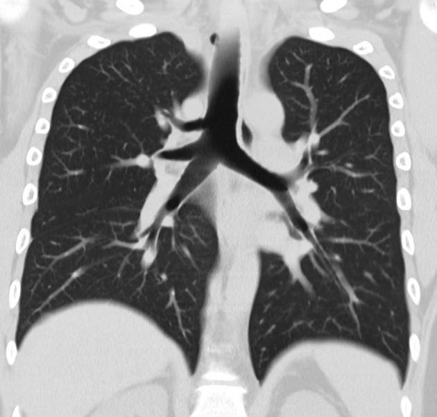 (
Image Source
)
Questions
- Describe the key anatomical abnormality demonstrated on the chest computed tomography scan.
- Differentiate between a "displaced" (ectopic) tracheal bronchus and a "supernumerary" tracheal bronchus.
- List three acute perioperative or anesthetic complications associated with the presence of this anomaly during endotracheal intubation.
- Outline the conservative management strategies and the definitive surgical indications for this congenital malformation.
Answer
- CT Chest Finding:
- Aberrant tracheal bronchus (bronchus suis / pig bronchus): An anomalous ectopic bronchus originating directly from the lateral wall of the trachea proximal to the main carina (usually within 2 cm above carina on the right side), aerating the apical or entire right upper lobe.
- Displaced vs Supernumerary Tracheal Bronchus:
- Displaced (Ectopic) Type: The normal right upper lobe bronchus (or one of its segmental branches, typically the apical segment B1) is missing from the right mainstem bronchus and instead takes origin aberrantly from the trachea. The total number of segmental bronchi is conserved.
- Supernumerary Type: The right mainstem bronchus bifurcates/trifurcates normally with all physiological segmental bronchi present; an additional, accessory anomalous bronchus originates from the trachea to supply redundant lung parenchyma or a normal segment.
- Anesthetic and Perioperative Complications:
- Obstruction of the tracheal bronchus: An endotracheal tube (ETT) cuff or tip placed distal to the aberrant orifice occludes it, causing acute right upper lobe atelectasis, retention of secretions, and severe hypoxemia.
- Accidental intubation of the tracheal bronchus: The ETT may selectively cannulate the anomalous bronchus, causing unilateral single-segment ventilation, failure of whole-lung ventilation, and tension pneumothorax.
- Ineffective one-lung ventilation: Inability to isolate the right lung during thoracic surgery using standard double-lumen tubes or bronchial blockers.
- Management:
- Conservative Management: Indicated in asymptomatic or minimally symptomatic individuals: Chest physiotherapy, postural drainage, prompt broad-spectrum antimicrobial coverage for acute infections, and surveillance.
- Definitive Surgical Management: Indicated in patients with recurrent pneumonias, persistent lobar atelectasis, localized bronchiectasis, hemoptysis, or associated congenital cystic adenomatoid malformation:
- Surgical resection (segmentectomy or right upper lobectomy via video-assisted thoracoscopic surgery [VATS] or thoracotomy).
- Bronchoplastic reimplantation into the right mainstem bronchus (rarely indicated).
More Details
RES066 - Recurrent Lobar Infiltrate in Toddler
Scenario
A 28-month-old previously thriving boy is brought to the pediatric outpatient department with high-grade fever and productive cough for 4 days. Medical records reveal that he was treated for right middle lobe consolidation with oral amoxicillin-clavulanate 3 weeks ago, with complete radiological resolution documented at 10 days post-treatment. Physical examination reveals a temperature of 38.8°C, respiratory rate of 46/min, heart rate of 128/min, and oxygen saturation ($SpO_2$) of 94% on room air. Auscultation reveals localized tubular bronchial breath sounds, coarse inspiratory crackles, and monophonic expiratory wheezing over the right anterior mammary and axillary regions. Mantoux tuberculin skin test placed 48 hours ago shows 0 mm induration. Repeat chest radiography demonstrates dense homogenous opacification of the right middle lobe with an air bronchogram.
Questions
- What is the most likely underlying diagnosis precipitating this recurrent lobar pneumonia?
- Enlist four other classic clinical or radiological presentations of this condition in toddlers.
- Detail the anatomical reasons why this pathology preferentially involves the right tracheobronchial tree over the left.
- Specify the definitive diagnostic and therapeutic procedure of choice, including the preferred instrument and immediate post-procedure pharmacological management.
Answer
- Most Likely Underlying Diagnosis:
- Tracheobronchial foreign body aspiration (retained organic or inorganic foreign body causing ball-valve or complete bronchial obstruction leading to secondary obstructive post-obstructive pneumonia).
- Clinical and Radiological Presentations:
- Acute choking episode / penetration syndrome: Sudden onset coughing, gagging, paroxysmal cyanosis, or stridor while eating or playing.
- Intractable unilateral monophonic wheeze: Localized wheezing refractory to inhaled short-acting beta-2 agonists and systemic corticosteroids.
- Check-valve hyperinflation: Unilateral or lobar air-trapping visible on expiratory chest radiography or decubitus views, causing ipsilateral mediastinal shift away from the affected side.
- Resorption atelectasis / volume loss: Complete stop-valve obstruction causing lobar or whole-lung collapse with ipsilateral mediastinal shift toward the affected side.
- Anatomical Predisposition for Right Main Bronchus:
- Wider diameter: The right mainstem bronchus is significantly wider in caliber than the left.
- More vertical alignment: The right main bronchus diverges from the carina at an angle of 20°–30° from the vertical mid-tracheal line, compared to 45°–55° for the left main bronchus.
- Direct anatomical continuation: The right mainstem bronchus lies more in direct axial alignment with the trachea, creating a straighter path for aspirated objects carried by inspiratory air currents.
- Carina position: The tracheal carina is situated slightly to the left of the midline, preferentially channeling airflow and debris into the right bronchial orifice.
- Definitive Procedure and Management:
- Procedure of choice: Rigid bronchoscopy under general anesthesia using optical forceps with ventilating barrel (gold standard for safe retrieval and airway maintenance).
- Post-procedure pharmacotherapy:
- Intravenous dexamethasone: 0.5 mg/kg/day divided q6h for 24–48 hours to mitigate post-instrumentation subglottic and bronchial edema.
- Chest physiotherapy and postural drainage initiated after airway clearance (contraindicated before bronchoscopy due to dislodgement risk).
- Empirical antibiotics targeting post-obstructive secondary infection: Intravenous ampicillin-sulbactam 150 mg/kg/day IV divided q6h OR amoxicillin-clavulanic acid 90 mg/kg/day (amoxicillin component) divided q8h for 7 to 10 days.
More Details
graph TD
    A[Aspirated Foreign Body Lodges in Bronchus] --> B{Obstruction Type}
    B -->|Bypass Valve| C[Minimal Airway Resistance -> Normal CXR]
    B -->|Check Valve / Ball Valve| D[Air Enters on Inspiration, Trapped on Expiration -> Hyperinflation & Air Trapping]
    B -->|Stop Valve| E[No Air Enters or Exits -> Absorption Atelectasis & Collapse]
    D --> F[Persistent Mucosal Edema & Granulation]
    E --> F
    F --> G[Secondary Post-Obstructive Bacterial Pneumonia]
    G --> H[Recurrent Infiltrates in Same Anatomical Lobe]
RES067 - Evaluation of Mycobacterial Skin Tests
Scenario
A 6-year-old girl with a documented history of BCG vaccination at birth presents for tuberculosis screening following exposure to her father, who was diagnosed with smear-positive pulmonary tuberculosis 2 weeks ago. The child is entirely asymptomatic, with normal anthropometry (weight: 20 kg, $Z$-score: 0) and a normal chest radiograph. A newer specific intradermal skin test (Cy-Tb / C-Tb test) is being considered alongside traditional Purified Protein Derivative (PPD) Mantoux testing.
Questions
- Define the Cy-Tb (C-Tb) skin test and explain how it combines the operational advantages of traditional tuberculin skin testing (TST) with the immunological specificity of interferon-gamma release assays (IGRAs).
- Name the specific recombinant Mycobacterium tuberculosis antigens utilized in the C-Tb formulation and state their genomic encoding region.
- State the universal diagnostic cutoff threshold for induration positivity of the C-Tb test, including in immunocompromised individuals.
- Compare the performance of the C-Tb test against standard PPD TST with respect to confounding by prior BCG vaccination and non-tuberculous mycobacterial (NTM) sensitization.
Answer
- Definition and Operational Principle of Cy-Tb / C-Tb:
- The Cy-Tb (C-Tb) test is a novel, specific intradermal mycobacterial skin test formulated with recombinant Mycobacterium tuberculosis antigens.
- Combination of benefits: It retains the field simplicity, low cost, and lack of phlebotomy or specialized laboratory infrastructure characteristic of the standard TST, while achieving the high diagnostic specificity (>97%) of laboratory-based IGRAs by eliminating cross-reactivity with environmental mycobacteria and BCG.
- Antigens and Genetic Region:
- Antigens: Early Secretory Antigenic Target 6 kDa (ESAT-6) and Culture Filtrate Protein 10 kDa (CFP-10).
- Genomic Region: Region of Difference 1 (RD1), which is present in virulent M. tuberculosis and M. bovis, but completely deleted from all substrains of Bacillus Calmette-Guérin (BCG) and absent in most non-tuberculous mycobacteria (except M. marinum, M. kansasii, and M. szulgai).
- Diagnostic Positivity Cutoff:
- Universal cutoff: Transverse induration $\ge 5\text{ mm}$ measured at 48 to 72 hours via the standard ballpoint pen or caliper palpation technique.
- Unlike standard TST (which uses stratified cutoffs of 5 mm, 10 mm, and 15 mm), C-Tb utilizes a single universal cutoff of $\ge 5\text{ mm}$, regardless of HIV status, nutritional state, or age.
- Comparison with Standard PPD TST:
- BCG Cross-Reactivity: Standard PPD TST cross-reacts heavily with BCG strains, leading to false-positive indurations (especially within 2–5 years of vaccination). C-Tb antigens (ESAT-6 and CFP-10) are absent in all BCG strains; hence, C-Tb is not affected by prior BCG vaccination.
- Non-Tuberculous Mycobacteria (NTM): PPD contains over 200 shared mycobacterial antigens, causing widespread false-positive tests in NTM-endemic areas. C-Tb does not cross-react with environmental NTMs (such as M. avium complex), drastically reducing false positives.
RES068 - Asymptomatic Positive Intradermal Tuberculin Reaction
Scenario
A 3-year-old boy weighing 14 kg is evaluated at the pediatric chest clinic after his grandmother was diagnosed with microbiologically confirmed multidrug-susceptible pulmonary tuberculosis (rifampicin-sensitive, isoniazid-sensitive). The child is clinically asymptomatic with normal vital signs, unlabored respiration, and no lymphadenopathy. His weight-for-age is at the $-1.2$ $Z$-score. A Mantoux tuberculin skin test (2 TU PPD RT23) reveals an induration of 14 mm at 72 hours. Digital chest radiography (posteroanterior view) is completely normal. Sputum/gastric aspirate samples are not collected as the child has no clinical features or radiographic signs of disease.
Questions
- Define "Tuberculosis Infection" (TBI) in accordance with World Health Organization (WHO) and National Tuberculosis Elimination Program (NTEP) definitions.
- Outline the anthropometric criteria defining underweight and severe wasting in pediatric tuberculosis suspect evaluations for children $<10$ years and adolescents.
- List the three approved Tuberculosis Preventive Treatment (TPT) drug regimens recommended by WHO/NTEP for this child.
- Calculate the precise daily/weekly drug dosages, frequencies, and durations for the 3HR and 3HP regimens for this 14 kg child.
Answer
- Definition of Tuberculosis Infection (TBI):
- A state of persistent immune response to stimulation by Mycobacterium tuberculosis antigens with immunological evidence of sensitization (positive TST or IGRA) without evidence of clinically manifest, active tuberculosis disease (as ruled out by absence of symptoms, normal physical examination, and negative chest imaging).
- Anthropometric Cutoffs for Underweight in TB Evaluation:
- Children $<10$ years of age:
- Underweight: Weight-for-age $< -2$ $Z$-scores (SD).
- Severe Acute Malnutrition (Severe Wasting): Weight-for-height / length $< -3$ $Z$-scores (SD) or bilateral nutritional pitting edema or mid-upper arm circumference (MUAC) $< 115\text{ mm}$ (for 6–59 months).
- Adolescents ($\ge 10$ to 19 years) and adults:
- Body Mass Index (BMI) $< 18.5\text{ kg/m}^2$ (or BMI-for-age $< -2$ $Z$-score per WHO pediatric growth charts).
- Children $<10$ years of age:
- Approved TPT Regimens:
- 3HR: 3 months of daily Isoniazid plus Rifampicin.
- 3HP: 3 months of once-weekly Isoniazid plus Rifapentine (approved for children $\ge 2$ years of age).
- 6H: 6 months of daily Isoniazid monotherapy.
- Calculations and Regimen Dosing for a 14 kg Child:
- Regimen 1: 3HR (Daily Rifampicin + Isoniazid for 3 months / 84 doses):
- Isoniazid (H): $10\text{ mg/kg/day}$ (Range: 7–15 mg/kg/day) $$\text{Dose} = 14\text{ kg} \times 10\text{ mg/kg/day} = \mathbf{140\text{ mg/day}} \quad (\text{Target: } 100\text{--}150\text{ mg/day}) $$
- Rifampicin (R): $15\text{ mg/kg/day}$ (Range: 10–20 mg/kg/day) $$\text{Dose} = 14\text{ kg} \times 15\text{ mg/kg/day} = \mathbf{210\text{ mg/day}} \quad (\text{Target: } 150\text{--}250\text{ mg/day}) $$
- Or fixed-dose dispersible tablets: 3 tablets daily of pediatric FDC containing (Rifampicin 75 mg + Isoniazid 50 mg) for 14 kg body weight (12–15 kg weight band: 3 tablets).
- Pyridoxine (Vitamin B6): 10 mg/day orally co-administered.
- Regimen 2: 3HP (Once-weekly Rifapentine + Isoniazid for 12 weeks / 12 doses):
- For weight band 10.0–15.9 kg:
- Isoniazid: $500\text{ mg}$ orally once weekly.
- Rifapentine: $300\text{ mg}$ orally once weekly.
- Total: 12 weekly doses taken with food.
- For weight band 10.0–15.9 kg:
- Regimen 1: 3HR (Daily Rifampicin + Isoniazid for 3 months / 84 doses):
RES069 - Abnormal Mechanical Ventilator Pressure Loops
Scenario
A 4-year-old girl (weight: 16 kg) with severe bacterial meningitis and Glasgow Coma Scale (GCS) score of 6 is intubated with an uncuffed endotracheal tube and mechanically ventilated in the Pediatric Intensive Care Unit (PICU). The ventilator mode is Volume-Controlled Continuous Mandatory Ventilation (VC-CMV) with a set tidal volume of 128 mL ($8\text{ mL/kg}$), positive end-expiratory pressure (PEEP) of $5\text{ cmH}_2\text{O}$, and inspiratory time of 0.7 seconds. During rounds, the ventilator displays the following real-time pressure-volume (P-V) loop and tidal volume scalar:
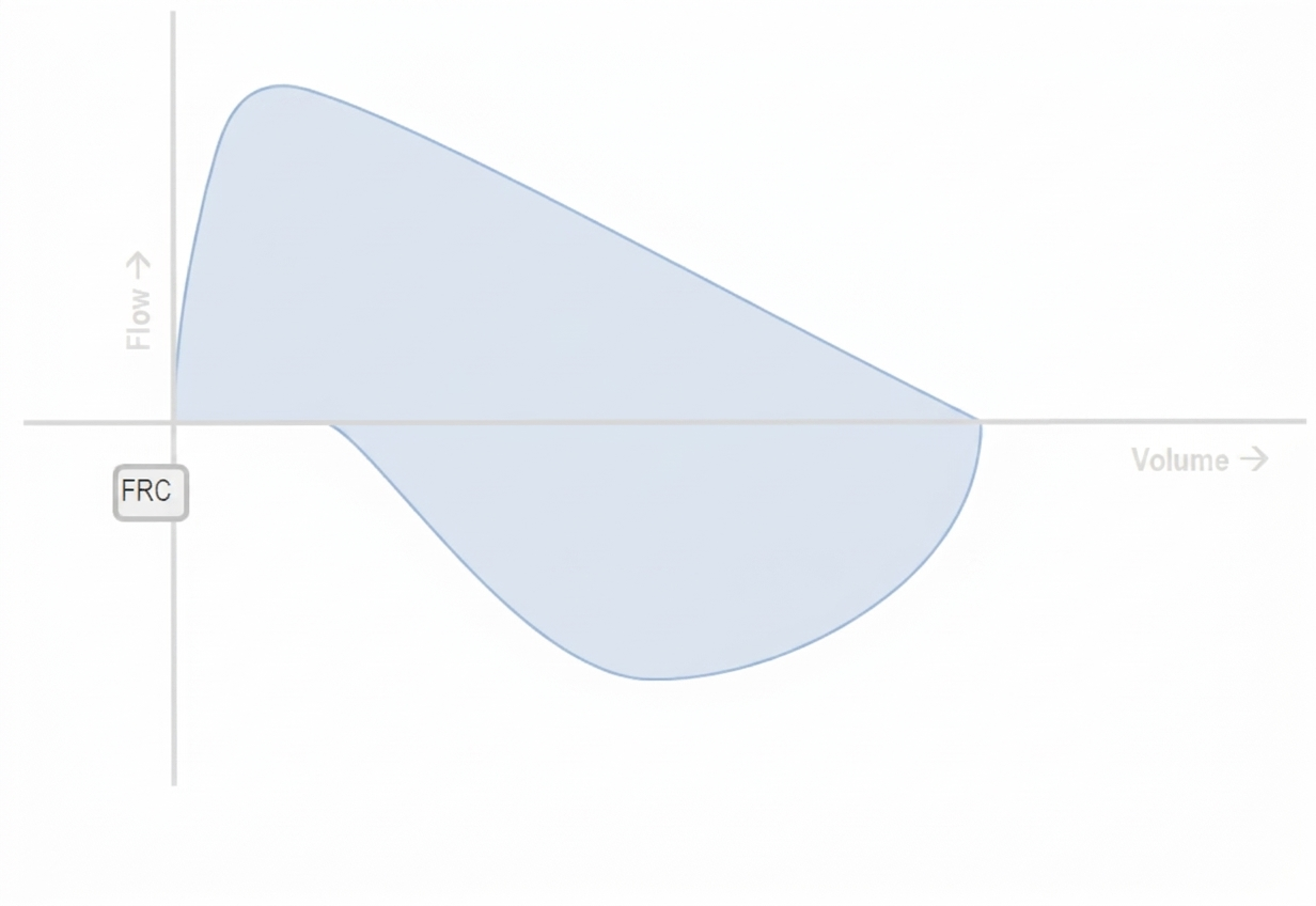
Questions
- Describe the pathognomonic graphic abnormality displayed on the Pressure-Volume loop.
- What underlying mechanical ventilatory complication is depicted?
- Enlist four distinct potential causes of this finding, categorizing them into patient/airway-related and circuit-related factors.
- If the recorded delivered inspiratory tidal volume ($V_{Ti}$) is 180 mL and the measured exhaled expiratory tidal volume ($V_{Te}$) returned to the exhalation block is 126 mL, calculate the absolute leak volume and the percentage air leak.
Answer
- Graphic Abnormality on the Pressure-Volume Loop:
- The expiratory limb of the pressure-volume loop does not return to the volume baseline (zero or PEEP point) at end-expiration.
- The loop fails to close, creating an open-ended loop or "gap" between inspiratory and expiratory volume curves at the conclusion of exhalation.
- Mechanical Ventilatory Complication:
- Air leak / Circuit-Airway volume mismatch (Expiratory tidal volume significantly lower than inspiratory tidal volume).
- Causes of Ventilator Circuit / Airway Leak:
- Patient / Airway-Related Causes:
- Inappropriate endotracheal tube (ETT) size (undersized uncuffed ETT with excessive peritubular glottic leak).
- Deflated, punctured, or ruptured ETT cuff (in cuffed tubes).
- Malposition of ETT (cuff positioned at or above vocal cords / accidental partial extubation).
- Development of bronchopleural fistula with chest tube suction air evacuation.
- Circuit-Related Causes:
- Inadvertent disconnection or loose junction at the Y-piece, flow sensor, or humidifier chamber.
- Incompetent exhalation valve or defective exhalation membrane/cassette.
- Open, loose, or cracked water trap seal or inline nebulizer port.
- Patient / Airway-Related Causes:
- Mathematical Derivation of Leak: $$\begin{aligned} \text{Absolute Leak Volume} &= V_{Ti} - V_{Te} \\ &= 180\text{ mL} - 126\text{ mL} \\ &= \mathbf{54\text{ mL}} \end{aligned} $$ $$\begin{aligned} \text{Percentage Air Leak (\%)} &= \left( \frac{V_{Ti} - V_{Te}}{V_{Ti}} \right) \times 100 \\ &= \left( \frac{54\text{ mL}}{180\text{ mL}} \right) \times 100 \\ &= \mathbf{30\%} \quad (\text{Significant if } > 10\text{--}15\%) \end{aligned} $$
RES070 - Pediatric Upper Airway Flow Limitation
Scenario
Laryngeal and upper airway disorders in pediatric practice produce distinctive functional and anatomical derangements. Match the clinical vignettes described below with the appropriate endoscopic and clinical diagnoses:
Clinical Vignettes:
- Vignette 1: A 14-year-old competitive swimmer with diagnosed asthma presents with recurrent exercise-triggered breathlessness, throat tightness, and inspiratory wheeze. Symptoms peak abruptly during intense exertion and resolve completely within 5–10 minutes of stopping. High-dose inhaled corticosteroids and beta-agonists produce no relief.
- Vignette 2: A 3-week-old female infant who underwent surgical ductal clipping for persistent patent ductus arteriosus (PDA) 10 days ago presents with a weak, hoarse cry, frequent choking during breastfeeds, and mild biphasic stridor.
- Vignette 3: A 3-year-old boy presents with progressive dysphonia and hoarseness of 6 months' duration, now accompanied by progressive nocturnal inspiratory stridor. Flexible laryngoscopy reveals multiple pink, friable, raspberry-like exophytic papillomatous masses lining both true vocal folds.
- Vignette 4: A 6-week-old male infant presents with high-pitched inspiratory stridor that began at 2 weeks of age. The stridor intensifies when crying, agitated, or placed supine, and improves distinctly in the prone position. Feedings are well-tolerated and growth is normal.
Questions
- Provide the specific clinical diagnosis for each vignette (Vignette 1 to 4).
- For Vignette 1, describe the characteristic finding on spirometric maximal expiratory-inspiratory flow-volume loop and state the gold standard diagnostic investigation.
- For Vignette 2, identify the injured nerve, explain its specific anatomical vulnerability during PDA ligation, and name the resulting vocal cord resting position.
- For Vignette 3, identify the causative pathogen and primary first-line surgical management.
Answer
- Matching Diagnoses:
- Vignette 1: Vocal Cord Dysfunction (VCD) / Inducible Laryngeal Obstruction (ILO) / Paradoxical Vocal Fold Motion (PVFM).
- Vignette 2: Unilateral Left Vocal Cord Paralysis (secondary to Left Recurrent Laryngeal Nerve injury).
- Vignette 3: Recurrent Respiratory Papillomatosis (RRP) / Juvenile-Onset Laryngeal Papillomatosis.
- Vignette 4: Laryngomalacia (Congenital flaccid larynx).
- Evaluation of Vignette 1 (VCD / ILO):
- Flow-Volume Loop: Truncation / flattening of the inspiratory limb with a normal expiratory limb, demonstrating a variable extrathoracic upper airway obstruction pattern (Ratio of $FEF_{50\%} / FIF_{50\%} > 1.0$).
- Gold Standard Investigation: Continuous dynamic flexible laryngoscopy performed during exercise challenge (or symptom provocation), demonstrating paradoxical adduction of the anterior two-thirds of the vocal cords with a posterior chink during inspiration.
- Evaluation of Vignette 2 (Post-PDA Ligation Paralysis):
- Nerve Injured: Left Recurrent Laryngeal Nerve (branch of Left Vagus, CN X).
- Anatomical Course/Vulnerability: The left recurrent laryngeal nerve loops around the aortic arch immediately posterior to the ligamentum arteriosum / ductus arteriosus before ascending in the tracheoesophageal groove; it is vulnerable to direct traction, surgical dissection, electrocautery, or vascular clip application during PDA ductal ligation.
- Resting Position: The paralyzed left vocal fold typically rests in the paramedian or intermediate (cadaveric) position, leaving an incomplete glottic seal during phonation and swallowing.
- Evaluation of Vignette 3 (Recurrent Respiratory Papillomatosis):
- Causative Pathogen: Human Papillomavirus (HPV), predominantly low-risk genotypes HPV 6 and HPV 11 (acquired via vertical transmission during passage through an infected birth canal).
- First-Line Surgical Management: Cold micro-laryngeal debridement using a microdebrider (laryngeal shaver) OR pulsed potassium titanyl phosphate (KTP) / pulsed dye laser / $CO_2$ laser debulking under suspension microlaryngoscopy to clear the airway while preserving native vocal fold architecture.
More Details
graph TD
    A[Pediatric Laryngeal Pathology] --> B[Inspiratory Stridor]
    A --> C[Dysphonia & Aspiration]
    A --> D[Exercise-Induced Throat Tightness]

    B --> E[Dynamic Collapse on Inspiration: Laryngomalacia]
    B --> F[Exophytic Mucosal Proliferations: HPV 6 & 11 Papillomatosis]

    C --> G[Paramedian Cord Post-PDA Ligamentum Dissection: Left RLN Palsy]

    D --> H[Variable Extrathoracic Obstruction: Vocal Cord Dysfunction / ILO]
    H --> I[Flattened Inspiratory Flow-Volume Loop]
RES071 - Bedside Pediatric Audiological Evaluation
Scenario
A 7-year-old child is referred to the pediatric outpatient department for evaluation of reduced hearing and poor scholastic performance following recurrent episodes of upper respiratory infections. In the audiological assessment room, bedside tuning fork examinations using a 512 Hz fork are performed on five children presenting with suspected auditory deficits.
| Child | Weber Test Lateralization | Rinne Test: Right Ear | Rinne Test: Left Ear |
|---|---|---|---|
| Child 1 | Lateralizes to Right | Air Conduction < Bone Conduction (Negative) | Air Conduction > Bone Conduction (Positive) |
| Child 2 | Lateralizes to Right | Air Conduction > Bone Conduction (Positive) | Air Conduction > Bone Conduction (Positive) |
| Child 3 | Midline / No lateralization | Air Conduction > Bone Conduction (Positive) | Air Conduction > Bone Conduction (Positive) |
| Child 4 | Lateralizes to Left | Air Conduction > Bone Conduction (Positive) | Air Conduction < Bone Conduction (Negative) |
| Child 5 | Lateralizes to Left | Air Conduction > Bone Conduction (Positive) | Air Conduction > Bone Conduction (Positive) |
Questions
- Provide the definitive audiological diagnosis for each of the five children based on the tuning fork test findings.
- State the ideal frequency of the tuning fork used for these bedside examinations and justify why forks of 128 Hz and 2048 Hz are avoided.
- Explain the biophysical mechanism of sound lateralization during the Weber test in: a. Unilateral conductive hearing loss b. Unilateral sensorineural hearing loss
- What is a "False-Negative Rinne" test, in which clinical scenario does it arise, and what bedside technique prevents this diagnostic error?
Answer
Audiological Diagnoses:
- Child 1: Right ear conductive hearing loss (CHL).
- Child 2: Left ear sensorineural hearing loss (SNHL).
- Child 3: Normal hearing (or symmetrical bilateral sensorineural hearing loss).
- Child 4: Left ear conductive hearing loss (CHL).
- Child 5: Right ear sensorineural hearing loss (SNHL).
Tuning Fork Frequency Rationale:
- Standard frequency: 512 Hz (provides optimal balance between tactile vibration sense and tone duration).
- Why 128 Hz is avoided: Produces prominent bone-conducted vibrotactile sensations (somatosensory feeling rather than auditory perception), leading to false-positive bone conduction.
- Why 2048 Hz is avoided: Tone decays too rapidly (short decay time), limiting adequate comparison time between bone and air conduction.
Biophysical Mechanism of Weber Lateralization:
- Unilateral Conductive Hearing Loss (CHL):
- Lateralizes to the affected ear.
- Loss of ambient environmental acoustic masking (masking effect) in the affected ear enhances perception of bone-conducted sound.
- Occlusion effect: The middle ear pathology impedes the normal retrograde escape of acoustic energy through the ossicular chain and tympanic membrane, routing more sound directly to the cochlea.
- Unilateral Sensorineural Hearing Loss (SNHL):
- Lateralizes to the unaffected (better) ear.
- The cochlea or cochlear nerve on the diseased side has elevated auditory thresholds and reduced neural transductive capacity, whereas the contralateral intact cochlea detects bone vibrations normally.
- Unilateral Conductive Hearing Loss (CHL):
False-Negative Rinne:
- Definition: A test where the patient reports hearing bone conduction better than air conduction (mimicking a "negative" Rinne / CHL), but the sound is actually perceived via transcranial bone transmission by the contralateral normal cochlea.
- Clinical Scenario: Severe to profound unilateral sensorineural hearing loss ("dead ear").
- Prevention / Bedside Maneuver: Mask the contralateral (non-test) ear using a Bárány noise box, continuous tragal rubbing, or an air-jet generator to suppress cross-hearing during bone conduction assessment.
More Details
graph TD
    A[Weber Test Lateralization] --> B[Lateralizes to One Side]
    A --> C[Central / No Lateralization]

    B --> D[Check Rinne in that Ear]
    D -->|BC > AC: Negative| E[Conductive Hearing Loss in that Ear]
    D -->|AC > BC: Positive| F[Sensorineural Hearing Loss in Contralateral Ear]

    C --> G[Check Bilateral Rinne]
    G -->|AC > BC Bilaterally| H[Normal Hearing or Symmetrical SNHL]
    G -->|BC > AC Bilaterally| I[Bilateral Conductive Hearing Loss]
Clinical Pitfalls in Pediatric Practice:
- Transcranial Transmission: The interaural attenuation for bone conduction is approximately 0 to 10 dB. Therefore, intense bone vibration in an ear with no cochlear function is readily perceived by the healthy opposite ear. Always apply contralateral masking when severe unilateral hearing loss is suspected.
- Optimal Fork Placement: The stem of the tuning fork during the Weber test should be firmly placed on the midline vertex, forehead, or upper incisors (best bone coupling). Avoid placing it over hair or soft tissue.